
Fig. 7
- ID
- ZDB-FIG-100525-36
- Publication
- Okuda et al., 2010 - B1 SOX coordinate cell specification with patterning and morphogenesis in the early zebrafish embryo
- Other Figures
- All Figure Page
- Back to All Figure Page
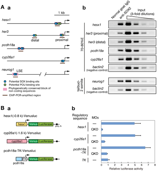
Direct regulatory targets of the B1 SOX proteins in the zebrafish embryo. (A) ChIP analysis showing direct association of B1 SOX with regulatory sequences of the downstream genes. (a) Potential binding sites for SOX and POU within the analyzed genomic regions are schematically shown. (b) ChIP-PCR analysis using anti-SOX2 antibody. ChIP experiments were performed using zebrafish embryos at the 70–80%E and tailbud to 2-somite stages. ChIP-PCR analysis using anti-SOX2 antibody that weakly cross-reacts with SOX3/19A/19B revealed specific binding of B1 SOX to the regulatory sequences of the hesx1, her3, pcdh18a, cyp26a1 and neurog1 genes in the zebrafish embryo. bactin2 was used as a negative control. (B) B1 SOX-dependent activities of the regulatory sequences of hesx1, cyp26a1 and pcdh18a. (a) The Venusluc fusion reporter (Venus plus firefly luciferase) constructs containing either of the promoters for hesx1 or cyp26a1 or the upstream conserved sequence of pcdh18a with the HSV TK promoter are schematically shown. (b) The Venusluc reporters were injected into embryos with or without the MOs for QKD together with the reference vector TK-Renilla luciferase. More than 20 injected embryos per sample were collected at the tailbud stage, and luciferase assays were performed. The normalized luciferase activity generated by TK-Venusluc was arbitrarily assigned a value of 1. Data are shown as the average values of four independent injection experiments with standard errors. |