
FIGURE
Fig. S6
- ID
- ZDB-FIG-100525-41
- Publication
- Okuda et al., 2010 - B1 SOX coordinate cell specification with patterning and morphogenesis in the early zebrafish embryo
- Other Figures
- All Figure Page
- Back to All Figure Page
Fig. S6
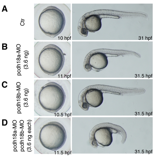
Phenotypes of pcdh18a and pcdh18b knockdowns. Bright-field images of live embryos at 10–11.5 hpf and 31–31.5 hpf are shown. (A) Uninjected control (Ctr) embryos. (B–D) Single and double knockdowns of pcdh18a/18b. Moderate amounts of MO (3.6 ng) were used for single KD (B,C) and a 1:1 mixture of two MOs (3.6 ng each) was injected for double KD (D). Double knockdown embryos showed a shorter anteroposterior axis than the single knockdown embryos, indicative of more severe defects in C&E movements and suggesting a degree of functional redundancy between pcdh18a and pcdh18b. |
Expression Data
Expression Detail
Antibody Labeling
Phenotype Data
Phenotype Detail
Acknowledgments
This image is the copyrighted work of the attributed author or publisher, and
ZFIN has permission only to display this image to its users.
Additional permissions should be obtained from the applicable author or publisher of the image.
Full text @ PLoS Genet.