|
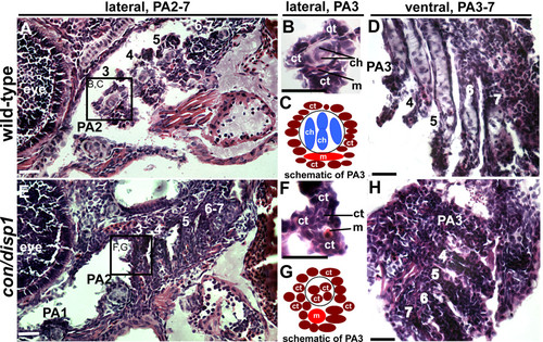
con/disp1 posterior arch-residing CNCC become fibrous-connective tissue. Lateral, (A,B,E,F) or ventral (D,H) H&E stained 10 μM sections of 5 dpf wild type (A,B,D) or con/disp1 mutant (E,F,H) larvae. (A,C) Chondrocytes are surrounded by connective tissue within each wild type PA (A), with higher magnification of PA3 revealing individual cell types (B). Box in (A) surrounding PA3 is viewed at higher magnification in (B). (E,F) con/disp1 mutants lack chondroctyes in PA3-7 (E), higher magnification of PA3 displays ectopic fibrous-connective tissue in PA3 (F). Box in (E) is viewed at higher magnification in (F). (C,G) Schematic of cell types visualized in wild type (C) and con/disp1 (G) PA3. (D,H) Horizontal sections show chondrocytes stacked within wild type posterior arches (D), while fibrous-connective tissue populate the con/disp1 posterior arches (H). Scale bar: 50 μM.
|