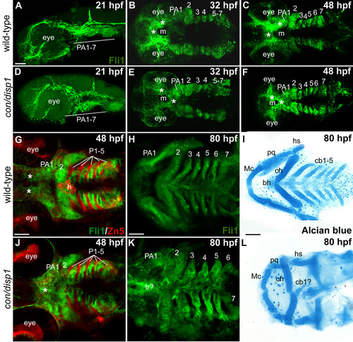
CNCC patterning in the con/disp1 mutant is visualized by the fli1gfp transgene. Lateral views (A,D) or ventral views (B,C,E-L) of confocal stack projections of fli1GFP+ CNCC in wild type and con/disp1 mutants (A-H, J,K) or Alcian blue-stained larvae to visualize cartilages (I,L). (G,J) Zn5 (red) staining to visualize endodermal pouches (P 1-5) in fli1GFP embryos. (A,D) At 21 hpf, wild type (A) and con/disp1 mutants (D) reveal similar patterns of postmigratory-CNCC. (B,C) 32 hpf ventral views of wild type embryos reveal CNC condensations in PA (B), and segmentation of CNCC by 48 hpf (C). (E,F) By 32 hpf, con/disp1 anterior-most CNCC (asterisks) become mispatterned, while CNCC within more PA 2-7 are correctly patterned (E) and segmented by 48 hpf (F). (G,J) CNCC in wild type (G) and con/disp1 mutants (J) are interdigitated properly in well-formed endodermal pouches 1-5 (P 1-5) (Zn5, red). (H,K) con/disp1 mutants show developed, segmented CNCC in posterior arches (K). (I,L) CNCC within con/disp1 mutant posterior arches fail to become cartilage. Scale bar: 50 μM.
|
