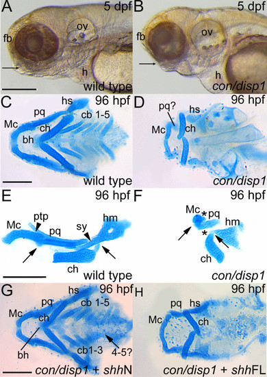
con/disp1 mutants display defective cartilage development in the PA which is partly rescued by RNA encoding non lipid-modified Shh. (A,B) Lateral views of live larvae show a reduction in head tissue and a deficit in jaw outgrowth (arrowhead) in con/disp1 mutant larvae at 5 dpf. (C) Ventral view of 96 hpf Alcian blue-stained wild type cartilages. (D) con/disp1 mutant cartilages reveal reduced, hypoplastic mandibular and hyoid arch cartilage elements and a complete absence of the cb 1-5 elements and midline-forming bh cartilage. (E,F) Lateral view of jaw cartilages reveals malformed joints (arrows denote joint site), fewer chondrocytes contributing to the pq cartilage element and a complete loss of the ptp and sy cartilage element in con/disp1 mutant (arrowhead denotes presence of cartilage, asterisk denotes cartilage absence). (G) Injection of 150 pg shhN mRNA into genotype-confirmed con/disp1 mutant mostly restores cartilage elements to wild type state (arrow indicates unilateral rescue in cb 4-5 elements), while (H) con/disp1 mutant injected with 150 pg shhFL mRNA are phenotypically similar to noninjected con/disp1 larvae. Scale bar: 50 μM.
|
