
Fig. S4
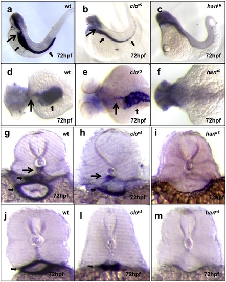
hands-off, but not cloche, mutant larvae show defective mural cell development. Seventy-two hpf larvae from wild-type crosses, or cloche (clos5)or hands-off (hans6)heterozygote incrosses analyzed for acta2 expression. In wild-type, acta2 expression identifies structures such as the dorsal aorta (long black arrows) and visceral smooth muscle (short black arrows). This expression is present in cloche mutants but missing in hands-off mutants. Images (a–c) are lateral views, anterior to the left; (d–f) dorsal views, anterior to the left; (g–i) cross-sections at the level of the 1st–2nd somite, and (j–m) cross-sections at the level of the 10–11th somite. |
| Gene: | |
|---|---|
| Fish: | |
| Anatomical Terms: | |
| Stage: | Protruding-mouth |
Reprinted from Mechanisms of Development, 126(8-9), Santoro, M.M., Pesce, G., and Stainier, D.Y., Characterization of vascular mural cells during zebrafish development, 638-649, Copyright (2009) with permission from Elsevier. Full text @ Mech. Dev.