
Fig. 7
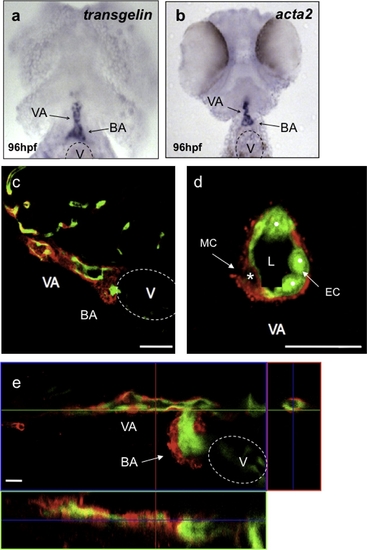
Vascular mural cells contribute to the ventral aorta and the bulbus arteriosus. Ninety-six hpf wild-type larvae analyzed for transgelin (a) and acta2 (b) expression. transgelin and acta2 are expressed in the bulbus arteriosus (BA) and ventral aorta (VA). (c) Confocal image of a sagittal section of a 96 hpf Tg(flk1:EGFP)s843 wild-type larva stained for Transgelin (red) show that vascular MCs surround the VA and contribute to the BA. (d) Confocal image of a transverse section of the VA shows single MCs around the ECs (white dots). (e) Confocal projection of the heart region of a 120 hpf Tg(flk1:EGFP)s843 wild-type larva stained for Transgelin (red). At this stage of development, vascular MCs are present in the BA as well as VA. Scale bar, 20 μm. V, ventricle; L, vascular lumen. |
| Genes: | |
|---|---|
| Antibody: | |
| Fish: | |
| Anatomical Terms: | |
| Stage Range: | Day 4 to Day 5 |
Reprinted from Mechanisms of Development, 126(8-9), Santoro, M.M., Pesce, G., and Stainier, D.Y., Characterization of vascular mural cells during zebrafish development, 638-649, Copyright (2009) with permission from Elsevier. Full text @ Mech. Dev.