
Fig. 3
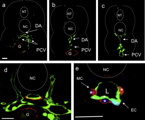
Vascular mural cells are present around the dorsal aorta in larvae. Confocal images of transverse sections of 80 hpf Tg(flk1:gfp)s843 wild-type larvae stained for Transgelin (red; (a–e)) and DNA (blue; (e)). Vascular MCs are Transgelin-positive cells surrounding endothelial cells (ECs) (green) of the dorsal aorta (DA). Sections shown are at the level of the 2nd (a, d and e), 10th (b) and 18th (c) somite. Vascular Transgelin-positive MCs are absent from the PCV in all sections (a–d). High-magnification images show that Transgelin-positive MCs (white asterisks) are single cells clearly distinguishable from endothelial cells (ECs: white dots). Scale bars, 20 μm. NT, neural tube; NC, notochord; DA, dorsal aorta; PCV, posterior cardinal vein; G, gut; L, vascular lumen. |
| Gene: | |
|---|---|
| Antibody: | |
| Fish: | |
| Anatomical Terms: | |
| Stage: | Protruding-mouth |
Reprinted from Mechanisms of Development, 126(8-9), Santoro, M.M., Pesce, G., and Stainier, D.Y., Characterization of vascular mural cells during zebrafish development, 638-649, Copyright (2009) with permission from Elsevier. Full text @ Mech. Dev.