
FIGURE
Fig. S1
Fig. S1
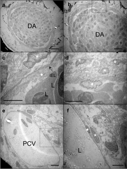
Transmission electron microscopy (TEM) images of 20 dpf zebrafish. (a and b) Sections of the DA at different magnifications, (c and d) higher magnification images of the area boxed in (b). (c) IEL is evident between EC layer (dot) and MCs (asterisk); (d) three layers of MCs are present around the EC layer, (e and f) section of the posterior cardinal vein (PCV) at high magnification, (f) higher magnification of EC (dot) and MC (asterisk) boxed in (e). Only a single layer of MCs is evident around the PCV. L, lumen; IEL, internal elastic lamina. Scale bars: 10 μm. |
Expression Data
Expression Detail
Antibody Labeling
Phenotype Data
Phenotype Detail
Acknowledgments
This image is the copyrighted work of the attributed author or publisher, and
ZFIN has permission only to display this image to its users.
Additional permissions should be obtained from the applicable author or publisher of the image.
Reprinted from Mechanisms of Development, 126(8-9), Santoro, M.M., Pesce, G., and Stainier, D.Y., Characterization of vascular mural cells during zebrafish development, 638-649, Copyright (2009) with permission from Elsevier. Full text @ Mech. Dev.