
FIGURE
Fig. 4
Fig. 4
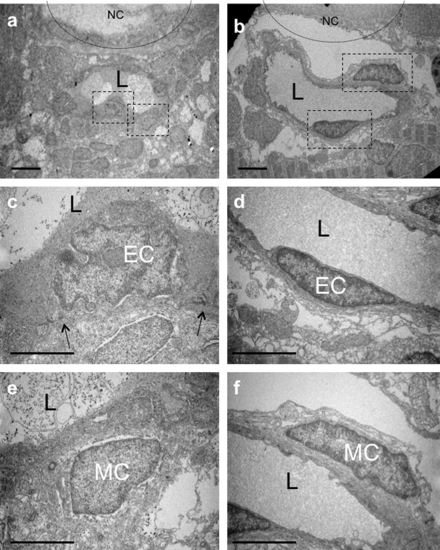
TEM of single mural cells surrounding endothelial vessels in larvae. Transmission electron microscopy (TEM) images of transverse sections of 80 (a, c and e) and 120 (b, d and f) hpf larvae. (c and e) Higher magnification of MCs and ECs boxed in (a). MCs and ECs exhibit different morphologies, and tight junctions are evident between ECs (black arrows). (d–f) Higher magnification images of MCs and ECs boxed in (b). Both ECs and MCs exhibit a more elongated morphology at 120 than 80 hpf. L, lumen. Scale bars, 5 μm. |
Expression Data
Expression Detail
Antibody Labeling
Phenotype Data
Phenotype Detail
Acknowledgments
This image is the copyrighted work of the attributed author or publisher, and
ZFIN has permission only to display this image to its users.
Additional permissions should be obtained from the applicable author or publisher of the image.
Reprinted from Mechanisms of Development, 126(8-9), Santoro, M.M., Pesce, G., and Stainier, D.Y., Characterization of vascular mural cells during zebrafish development, 638-649, Copyright (2009) with permission from Elsevier. Full text @ Mech. Dev.