
Fig. S6
- ID
- ZDB-FIG-090717-31
- Publication
- Winata et al., 2009 - Development of zebrafish swimbladder: the requirement of Hedgehog signaling in specification and organization of the three tissue layers
- Other Figures
- All Figure Page
- Back to All Figure Page
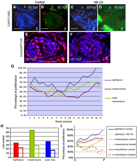
Analyses of apoptosis and proliferation in CA-treated embryos. (A–D) Detection of apoptotic cells using TUNEL labeling (B, D) on the background of DAPI staining (A, C). Swimbladder regions are denoted in red line. No difference in apoptosis could be observed between control (A,B) and CA-treated larvae (C,D). (E–F) Proliferation assay using PCNA antibody. Note the difference in swimbladder size of control (E) and CA-treated (F) embryos. Cell nuclei are visualized with Hoechst staining, proliferating cells are indicated by red fluorescent signal. (G) Proliferation levels in serial sections across the wild type zebrafish swimbladder from the anterior to posterior. (H) Comparison of cell numbers in swimbladders of control and CA-treated larvae. All three tissue layers showed significant reduction of total cell numbers in CA-treated larvae, but the largest reduction was seen in the mesenchyme, followed by outer mesothelium. Solid coloured bars represent control, while others represent CA-treated group. (I) Proliferation profile in control and CA-treated swimbladder in the posterior region. X-axis represents consecutive sections from anterior (A) to posterior (P) for the eight posteriormost sections that have significant difference in proliferation levels. Note the downward shift of proliferation levels in CA-treated larvae compared to control in each tissue layer. Abbreviations: g, gut; n, notochord; sb, swimbladder. Scale bars: 100 μm. |
Reprinted from Developmental Biology, 331(2), Winata, C.L., Korzh, S., Kondrychyn, I., Zheng, W., Korzh, V., and Gong, Z., Development of zebrafish swimbladder: the requirement of Hedgehog signaling in specification and organization of the three tissue layers, 222-236, Copyright (2009) with permission from Elsevier. Full text @ Dev. Biol.