
Fig. 5
- ID
- ZDB-FIG-090717-26
- Publication
- Winata et al., 2009 - Development of zebrafish swimbladder: the requirement of Hedgehog signaling in specification and organization of the three tissue layers
- Other Figures
- All Figure Page
- Back to All Figure Page
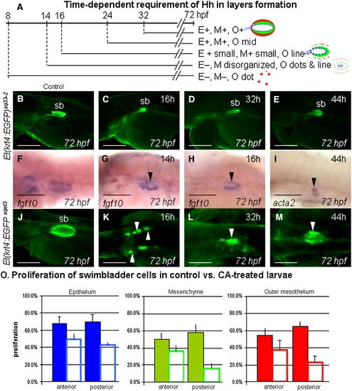
Stage-specific inhibition of Hh signaling by cyclopamine (CA). (A) Schematic representation of CA treatment experiments and observations on swimbladder development. CA treatment was performed starting from different developmental stages. Treatment results are summarized on the right. E, M and O indicate epithelium, mesenchyme and outer mesothelium respectively. “+” indicates presence and “-” indicates absence. 20 mM CA was used for early stage of treatment (16 hpf and earlier) and 50 mM and 100 mM CA was used for late stage of treatment (32 hpf and 44 hpf, respectively). (B–E) Expression of EGFP in swimbladder epithelium in Et(krt4:EGFP)sq33-2 embryos at 72 hpf after CA treatment stating at different time as indicated in the top-right of each panel. Panel (B) shows a control Et(krt4:EGFP)sq33-2 embryo without CA treatment. Note the hypomorphic epithelium in all treated larvae. (F–H) Mesenchymal fgf10 expression in control (F) and CA-treated larvae. Mesenchyme is present in larvae treated after 14 hpf (G), and is properly organized in larvae treated starting from 16 hpf (H). (I) Presence of acta2 expression in embryos treated with CA initiated at 44 hpf. (J–M) expression of EGFP in the outer mesothelium of control (J) and CA-treated Et(krt4:EGFP)sqet3 embryos (K–M). Note two lateral clusters of EGFP positive cells in 16 h treatment group, and partial organization in 32 h treatment group (white arrowhead). (O) Comparison of proliferation levels in control and CA-treated swimbladder showed a significant reduction in mesenchymal and outer mesothelial cell proliferation. Solid coloured bars represent control, while the open bars represent CA-treated samples. Proliferation percentage was observed in five anterior and posteriormost sections in each sample (for detail, see Supplementary Fig. 6 and legend). Abbreviations: sb, swimbladder. Scale bars: 250 μm. |
| Genes: | |
|---|---|
| Fish: | |
| Condition: | |
| Anatomical Terms: | |
| Stage: | Protruding-mouth |
Reprinted from Developmental Biology, 331(2), Winata, C.L., Korzh, S., Kondrychyn, I., Zheng, W., Korzh, V., and Gong, Z., Development of zebrafish swimbladder: the requirement of Hedgehog signaling in specification and organization of the three tissue layers, 222-236, Copyright (2009) with permission from Elsevier. Full text @ Dev. Biol.