
Fig. S4
- ID
- ZDB-FIG-090717-29
- Publication
- Winata et al., 2009 - Development of zebrafish swimbladder: the requirement of Hedgehog signaling in specification and organization of the three tissue layers
- Other Figures
- All Figure Page
- Back to All Figure Page
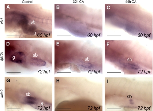
Expression of ptc1, fgf10a and acta2 in the swimbladder after CA inhibition of Hs signaling at late stages. (A–C) Expression of ptc1 in swimbladder of control and CA-treated larvae. Late stage CA treatments were performed starting from 32 hpf using 100 μM CA and from 44 hpf using 200 μM CA, which resulted in the elimination of ptc1 expression in the midline and in most of the visceral region. (D–F) fgf10a expression in the mesenchyme of swimbladder is present as normal. (G–I) Expression of acta2 is absent in 32 h CA-treated group, while present in small amount in 44 h CA-treated group, signifying a late requirement for Hh signaling in smooth muscle differentiation. Abbreviations: g, gut; sb, swimbladder. Scale bars: 250 μm. |
Reprinted from Developmental Biology, 331(2), Winata, C.L., Korzh, S., Kondrychyn, I., Zheng, W., Korzh, V., and Gong, Z., Development of zebrafish swimbladder: the requirement of Hedgehog signaling in specification and organization of the three tissue layers, 222-236, Copyright (2009) with permission from Elsevier. Full text @ Dev. Biol.