
Fig. 5
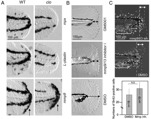
Role of Mmp9 expressed by non-myeloid cells during regeneration. (A) Normal expression of mmp9 in clo mutants, in which the myeloid cell differentiation is impaired. The expressions of mpx (top row), L-plastin (middle row) and mmp9 (bottom row) were detected in wild type larvae and clo mutants at 6 hpa. No significant decrease was observed for mmp9 expression (arrows), whereas the expression of mpx and L-plastin was undetectable (n = 8, respectively). The gene expression was confirmed by using 7–9 mutant larvae for the respective probes. (B) Abnormal larval regeneration caused by inhibitors of Mmps. Larvae exposed to GM6001 (0.1 mM, top) and Mmp9/13 inhibitor I (0.1 mM, middle) showed a deformed and irregular regeneration (9/9 for GM6001, and 19/32 for Mmp9/13 inhibitor I), whereas the amputated larvae exposed to the vehicle did not display any defect (bottom). Similar inhibitory effect was also observed using Mmp9-specifc inhibitor I (0.1 mM, 10/13, data not shown). The same magnifications for respective panels in A or B (scale bars in the top row). (C) Normal activation of cell proliferation in the presence of Mmp9 inhibitor. BrdU labeling was carried out 18–24 hpa in the presence (top) and absence (middle) of Mmp9 inhibitor. Significant difference of cell proliferation was not detected. Many of responding cells to amputation are present in areas indicated by double-headed arrows. The bracketed areas represent cells that fate to form the adult tail fins. The same magnifications for top and middle panels (scale bar in the top). Quantification (bottom) was carried out by counting BrdU-positive cells in the caudal fin fold (posterior to the dashed lines). Data in the graph are the mean ± SEM. Statistical significance was tested by Student's t-test at P = 0.01. N.S., not significant. |
| Genes: | |
|---|---|
| Fish: | |
| Condition: | |
| Anatomical Term: | |
| Stage: | Long-pec |
Reprinted from Developmental Biology, 325(1), Yoshinari, N., Ishida, T., Kudo, A., and Kawakami, A., Gene expression and functional analysis of zebrafish larval fin fold regeneration, 71-81, Copyright (2009) with permission from Elsevier. Full text @ Dev. Biol.