
Fig. S3
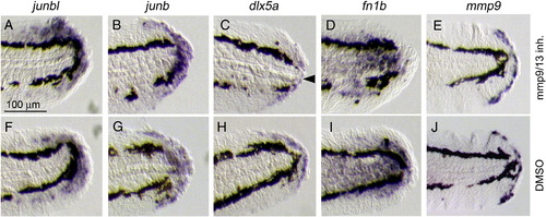
Formation of the blastema, wound epithelium and other regeneration-induced cell types in the presence of Mmp inhibitors. (A–J) ISH analysis for the induction of the respective genes after exposure to Mmp9/13 inhibitor I (A–E) or to the vehicle (F–J) at 1 dpa. The expressions of junbl (A, F), junb (B, G), dlx5a (C, H), fn1b (D, I) and mmp9 (E, J) were not abolished by the inhibitor, suggesting that the formation of respective cell groups in response to amputation was not impaired by the Mmp inhibitors. Note that a slight decrease of dlx5 expression (arrowhead), implying that the inhibitor could have an effect on the proper formation of wound epithelium. The gene expression was confirmed by using 7–10 drug-treated larvae for the respective probes. The same magnifications for all panels (scale bar in A). |
Reprinted from Developmental Biology, 325(1), Yoshinari, N., Ishida, T., Kudo, A., and Kawakami, A., Gene expression and functional analysis of zebrafish larval fin fold regeneration, 71-81, Copyright (2009) with permission from Elsevier. Full text @ Dev. Biol.