
Fig. S1
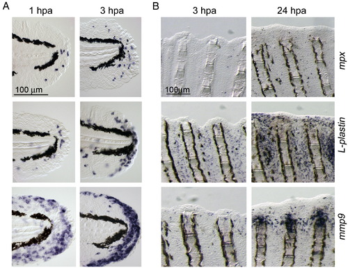
Distribution of neutrophils, macrophages, and mmp9-positive cells during larval and adult regeneration. (A) ISH analyses for expression of mpx, a neutrophils marker (top row), L-plastin, a macrophage marker (middle row) and mmp9 (bottom row) during larval fin fold regeneration. (B) Expression of mpx, L-plastin and mmp9 during adult fin regeneration. Only a few mpx-positive neutrophils were seen during regeneration up to 24 hpa (top row), whereas a large number of macrophages (middle row) were recruited to regions around the wound extending over 1 ray segment away from the stump. The mmp9-positive cells appeared by 24 hpa, however many mmp9-positive cells were localized at the amputation site and adjacent inter-ray regions with apparently different expression profile from mpx or L-plastin. The same magnifications for respective panels in A and B (scale bars in top left panels). |
Reprinted from Developmental Biology, 325(1), Yoshinari, N., Ishida, T., Kudo, A., and Kawakami, A., Gene expression and functional analysis of zebrafish larval fin fold regeneration, 71-81, Copyright (2009) with permission from Elsevier. Full text @ Dev. Biol.