
Fig. 4
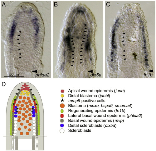
Cellular organization of regeneration niche in adult regenerates. (A–C) Large magnification of fin sections in which the expressions of phlda2 (A), dlx5a (B) and fn1b (C) were detected. phdla2 expression is seen at the lateral part of basal wound epidermis, with a gradient of weak expression to distal and proximal directions (A); whereas dlx5a expression shows a strong expression in cells proximal–lateral part of blastema in addition to a uniform weak expression in the basal wound epidermis. Judging from the continuous alignment of the large-sized cells (arrowheads) to the distal side of fin rays and from the coincidence with expressions of runx2a, runx2b, bmp6, bmp2b and ihha (Smith et al., 2006), these cells with strong dlx5a expression appear to be the early scleroblasts beginning to differentiate into the fin rays. The same magnifications for panels A–C (scale bar in panel A). (D) A schematic drawing showing the cellular organization in the adult fin regeneration niche. From the in situ expression analysis of regeneration-induced genes, at least 8 distinct cell types, 4 cell types in the wound epidermis, 3 in the blastema, and 1 unknown mmp9-positive cells, appear to be present during regeneration. |
| Genes: | |
|---|---|
| Fish: | |
| Condition: | |
| Anatomical Terms: | |
| Stage: | Adult |
Reprinted from Developmental Biology, 325(1), Yoshinari, N., Ishida, T., Kudo, A., and Kawakami, A., Gene expression and functional analysis of zebrafish larval fin fold regeneration, 71-81, Copyright (2009) with permission from Elsevier. Full text @ Dev. Biol.