|
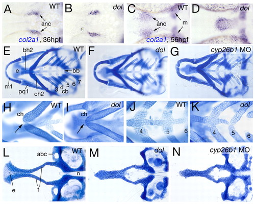
cyp26b1 mutants and morphants display deficiencies in midline cartilages of the neurocranium and visceral skeleton. All panels show ventral views of zebrafish head regions. (A-D) col2a1 in situ hybridization at indicated stages. (E-N) Alcian Blue stainings of cartilaginous craniofacial elements at 120 hpf. Pharyngeal arches are numbered (1, mandibulare; 2, hyoid; 3-7, branchial/gill arches 1-5). (E-G) Overviews of visceral skeleton. (H-K) Magnified views of ceratohyals (H,I) or pharyngeal arches 4-6 (J,K). Arrows in H,I point to ceratohyal (ch) attachment in midline. (L-N) Flat-mounts of neurocranium, revealing the absence of medial ethmoid (e) and anterior basicranial commissure (abc) in mutant and morphant. anc, chondrocytes of anterior neurocranium; bb, basibranchial; bh, basihyal; cb, ceratobranchials; m, Meckel's cartilage; n, notochord; pq, palatoquadrate; t, trabeculae cranii.
|