|
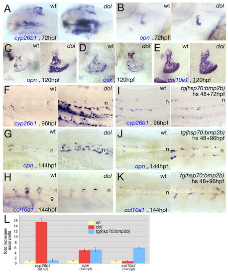
Loss of cyp26b1 and gain of Bmp signaling have different effects on the number and/or activity of osteoblasts. (A-K) In situ hybridization of zebrafish larvae of genotype indicated in upper right corners and with probes and at stages indicated at lower right corners. (A-I) Lateral views; (J,K) dorsal views. (A) Entire head; (B-E) opercle; (F-K) trunk at level of somites 6-10. In A, stronger cyp26b1 expression is seen in all craniofacial skeletogenic elements of the cyp26b1 mutant, but not in the dorsal brain. In G, perichordal opn-positive cells of the cyp26b1 mutant have largely given up their metameric distribution. (L) Average increase in the number of axial cyp26b1-(at 96 hpf), opn- or col10a1-positive cells (at 144 hpf) in the trunk/tail region of cyp26b1 mutants and heat-shocked Tg(hsp70:bmp2b) transgenic fish. Control wild-type (wt) siblings were set to a value of 1. Ten fish were counted per condition; standard errors are indicated. n, notochord.
|