
Fig. S5
- ID
- ZDB-FIG-081104-35
- Publication
- Laue et al., 2008 - Restriction of retinoic acid activity by Cyp26b1 is required for proper timing and patterning of osteogenesis during zebrafish development
- Other Figures
- All Figure Page
- Back to All Figure Page
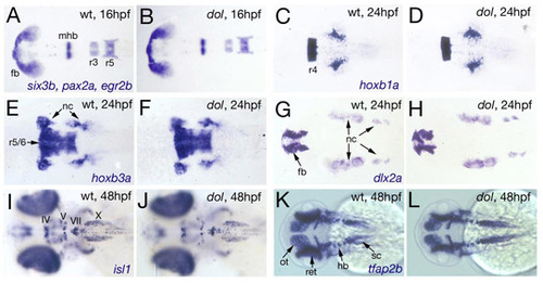
Brain patterning and cranial neural crest migration is unaffected in cyp26b1 mutants. (A-L) In situ hybridizations of cyp26b1 mutants (dol) and wild-type siblings (WT) at stages indicated in upper right corners, and with probes indicated in lower right corners; dorsal views on head regions. (A,B) Mutant shows normal expression patterns of six3b, a marker for forebrain (Kobayashi et al., 1998), pax2a, a marker for the midbrain-hindbrain boundary region (Krauss et al., 1991), and egr2b (formerly called krox20), a marker for hindbrain rhomobomeres 3 and 5 (Oxtoby and Jowett, 1993), indicating that brain patterning is unaffected, including normal sizing of the forebrain primordium. Similarly, expression patterns of hoxb1a, a marker for rhombomere 4 (Amores et al., 1998; Rohrschneider et al., 2007) (C,D), and hoxb3a, a marker for rhomobomeres 5 and 6 (Amores et al., 1998; Hogan et al., 2004) (E,F) are normal, indicating that cyp26b1 is dispensable for hindbrain patterning. Furthermore, cyp26b1 mutants display normal expression of hoxb3a in r5/r6-derived neural crest, and of dlx2b, a marker for all cranial neural crest cells (Akimenko et al., 1994), indicating that neural crest patterning and migration are unaffected by the mutation. Normal expression pattern of isl1 (Inoue et al., 1994) further indicates that branchiomotor neurons, derivatives of the hindbrain, are specified and patterned normally in cyp26b1 mutants (I,J). IV, trochlear nucleus; V, trigeminal nucleus; VII, facial nucleus; X, vagal nucleus). (K,L) Normal expression of tfap2b (Knight et al., 2005) in tectum, hindbrain and spinal cord of cyp26b1 mutant. fb, forebrain; hb, hindbrain; mhb, midbrain-hindbrain boundary; nc, neural crest; ot, optic tectum; r, rhomobomere; ret, retina; sc, spinal cord. |