
Fig. S3
- ID
- ZDB-FIG-081104-33
- Publication
- Laue et al., 2008 - Restriction of retinoic acid activity by Cyp26b1 is required for proper timing and patterning of osteogenesis during zebrafish development
- Other Figures
- All Figure Page
- Back to All Figure Page
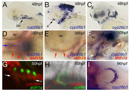
Expression pattern of cyp26b1 in comparison to cyp26a1, cyp26c1 and aldh1a, and in pectoral fin buds. (A-I) Wild-type zebrafish after whole-mount in situ hybridizations at stages indicated in upper right corners with probes indicated in lower right corners. (A-C,E) Lateral views; (D) ventral view; (F-I) ventrolateral views. (A-C) cyp26a1, cyp26b1 and cyp26c1 show distinct, but partially overlapping expression patterns. In B, unique expression of cyp26b1 in the condensation of the ethmoid plate is indicated by an arrow. (D-E) aldh1a is expressed adjacent to the eye vesicles (red arrows in D,E), lateral of the cyp26b1 expression domain in the midline of the developing neurocranium (blue arrow in D). (F,G) aldh1a is expressed in discrete domains lateral of the cyp26b1 expression domains in the branchial arches; the midline is indicated by arrowheads. Together, this points to the possible existence of a mediolateral RA gradient in the craniofacial system. (H,I) Similar to the earlier exclusion of cyp26b1 expression from chondrogenic neural crest cells of the craniofacial system (see Fig. 2), cyp26b1 is expressed in a distal crescent of the early pectoral fin bud, while the sox9a-positive chondrocyte precursors (Wada et al., 2005) are positioned more proximally (H). A corresponding distribution has been described for mouse Cyp26b1 in the developing limb buds (Yashiro et al., 2004), suggesting that similar proximodistal patterning processes might occur in both zebrafish and mouse, accounting for the profound limb defects in mouse Cyp26b1 mutants (Yashiro et al., 2004), and the moderate pectoral fin defects in zebrafish cyp26b1 mutants (see Fig. 1C,D). e, ethmoid plate; pf, pectoral fin; pp, pharyngeal pouches. |