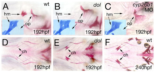
cyp26b1 mutants and morphants display increased ossification of endochondral and intramembranous craniofacial bones. (A-C) Lateral views and (D-F) ventral views of zebrafish larval heads after staining of ossified matrix with Alizarin Red at indicated ages. Insets in A-C show hyomandibula (hm; dorsal element of arch 2) of larvae of same genotype stained with Alcian Blue at 120 hpf. Mutant and morphant show an opercle (op) of increased size, whereas the hyomandibula fails to ossify, although its cartilage model is properly formed (insets). A similar combination of gain of opercle and loss of hyomandibula ossification has previously been described for endothelin mutants (Kimmel et al., 2003), possibly reflecting a morphogenetic effect of signaling to pattern ossification along the dorsoventral axis of the second arch and its associated elements. (D-F) Endochondral ossification within the ceratohyal (ch) is much more advanced in the mutant (E), comparable to the situation in a wild-type sibling 2 days later (F).
|
