
Fig. 4
- ID
- ZDB-FIG-080922-7
- Publication
- Guo et al., 1999 - Mutations in the zebrafish unmask shared regulatory pathways controlling the development of catecholaminergic neurons
- Other Figures
- All Figure Page
- Back to All Figure Page
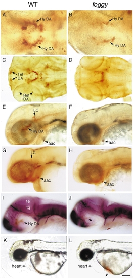
foggy mutant embryos have reduced DA neurons in the telencephalon, retina, and hypothalamus. Wildtype (left) and foggy mutant (right) embryos labeled with antibodies to (A–F) TH, (G and H) DβH, or (I and J) tubulin. (A and B) Ventral views of 2-day-old embryos showing reduced THir and number of hypothalamic DA neurons. (C and D) Ventral view of 3-day-old embryos showing the absence of THir telencephalic and retinal DA neurons. (E–H) Lateral views of 2-day-old embryos showing the absence of LC. (I and J) Lateral views of 36-h embryos showing normal axonal tracts. (K and L) 2-day-old live embryos depict accumulation of blood ventral to the heart due to lack of circulation. Abbreviations: aac, arch-associated CA cells; Hy DA, hypothalamic dopaminergic neurons; LC, locus coeruleus; te, tectum; tg, tegmentum; tel, telencephalon; ret, retina. Scale bar: 48 μm (A, B), 96 μm (C–J), and 192 μm (K, L). |
| Gene: | |
|---|---|
| Fish: | |
| Anatomical Terms: | |
| Stage Range: | Long-pec to Protruding-mouth |
| Fish: | |
|---|---|
| Observed In: | |
| Stage Range: | Prim-25 to Day 4 |
Reprinted from Developmental Biology, 208, Guo, S., Wilson, S.W., Cooke, S., Chitnis, A.B., Driever, W., and Rosenthal, A., Mutations in the zebrafish unmask shared regulatory pathways controlling the development of catecholaminergic neurons, 473-487, Copyright (1999) with permission from Elsevier. Full text @ Dev. Biol.