
Fig. 1
- ID
- ZDB-FIG-080922-4
- Publication
- Guo et al., 1999 - Mutations in the zebrafish unmask shared regulatory pathways controlling the development of catecholaminergic neurons
- Other Figures
- All Figure Page
- Back to All Figure Page
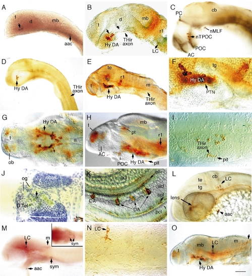
Catecholaminergic neurons in the brain of zebrafish embryos. Whole-mount embryos with rostral to the left (except I, J, and N), labeled with digoxygenin–RNA antisense probes to TH (A) and DβH (M) or antibodies to TH (rest of the images). (A) TH-positive neurons in a 20- to 23-somite embryo. (B) Hypothalamic DA neurons in a prim-12 (28 hpf) embryo are present rostral to the ventral flexure (indicated by an arrowhead) in the ventral diencephalon (the hypothalamus). Locus coeruleus (LC) neurons are present adjacent to rhombomere 1 (r1). (C) DA neurons are distributed along the TPOC (labeled in black with anti-acetylated tubulin antibody) in a prim-12 embryo. (D) Lateral view of a prim-12 embryo showing THir axons extending to the rostral spinal cord. (E–G) Lateral (E–F) and ventral (G) views of THir neurons in 50-h (E and F) and 72-h (G) embryos. (H–I) Lateral view and transverse section of the THir hypothalamus in a 96-h fry. Axonal labeling in the pituitary is indicated in I. (J) Cross section of THir telencephalon in a 96-h fry showing dopaminergic innervation of olfactory glomerular structures. (K) DA amacrine neurons in the retina of a 96-hpf fry. (L) THir LC neurons and arch-associated CA cells (aac) in a prim-15 embryo (30 h). (M) Labeling of a 48-h embryo with DβH RNA probe shows that LC, caudal hindbrain, arch-associated, and sympathetic neurons are noradrenergic. Inset depicts sympathetic neurons in the plane of focus. (N) Transverse section showing LC neuron in a 96-h fry with polarized axonal projection. (O) A 96-hpf fry showing THir neurons in the caudal medulla. Abbreviations: aac, arch-associated CA cells; AC, anterior commissure; acl, amacrine cell layer; cb, cerebellum; d, diencephalon; D.Tel, dorsal telencephalon; V.Tel, ventral telencephalon; gcl, ganglion cell layer; hy, hypothalamus; ipl, inner plexiform layer; LC, locus coeruleus; m, medulla; mb, midbrain; n, notochord; nMLF, nucleus of the medial longitudinal fascicle; nTPOC, nucleus of the tract of postoptic commisure; ob, olfactory bulb; og, olfactory glomerule; opl, outer plexiform layer; PC, posterior commisure; pcl, phororeceptor cell layer; pit, pituitary; POC, postoptic commisure; pt, pretectum; PTN, posterior tubercular nuclei; r1, rhombomere 1; sym, sympathetic neurons; t, telencephalon; te, tectum; tg, tegmentum. Scale bar: 90 μm (A, B, C, G, H, L), 180 μm (D, E, M, O), 45 μm (F, I, J, N), and 30 μm (K). |
Reprinted from Developmental Biology, 208, Guo, S., Wilson, S.W., Cooke, S., Chitnis, A.B., Driever, W., and Rosenthal, A., Mutations in the zebrafish unmask shared regulatory pathways controlling the development of catecholaminergic neurons, 473-487, Copyright (1999) with permission from Elsevier. Full text @ Dev. Biol.