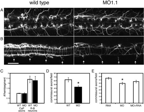
Pcdh1α splice-blocking morpholinos result in loss of neuronal subsets. (A) Immunolabeling of either wild type or pcdh1α-morpholino-injected embryos with a monoclonal antibody against acetylated tubulin prominently labeled neuronal cell bodies and axons, including Rohon–Beard cells, which are large sensory neurons present in the dorsal spinal cord. These cells extend elaborate sensory arbors, which innvervate the skin. Maximum intensity projections from optical sections of the dorsal spinal cord revealed the bilateral arrays of Rohon–Beard cells and a portion of their sensory arbors. Pcdh1α-morpholino-injection did not lead to any significant loss of Rohon–Beard cells, nor did it affect arborization of their sensory axons (C). (B) Maximum intensity projections of the ventral spinal cord revealed longitudinal axon tracts, as well as commissural axons. In addition, this view revealed the exit of CaP motor axons from the spinal cord at the ventral roots. In contrast to wild type embryos, pcdh1α-morpholino-injected embryos exhibited a loss of both longitudinal axons and commissural axons. However, there was no difference in the number of CaP motor axons. (C) As is apparent in (a) and b), there was no difference in either the number of CaP motoneurons or Rohon–Beard sensory neurons (32 spinal segments from 8 embryos for each data set). D, There was a substantial loss of commissural axons in pcdh1α-morpholino-injected embryos. Wild type embryos had an average of 62 ± 5.7 (mean ± SEM, n = 8 embryos) commissural axons within 4 spinal segments at 26 hpf. In a matched set of embryos injected with MO1.1, there were 42 ± 4.6 (mean ± SEM, n = 8 embryos) commissural axons per 4 spinal segments. This difference was found to be statistically significant (p < 0.05, Student's t-test). (E) Expression of the soluble Pcdhα cytoplasmic domain partially attenuates the loss of commissural axons. Embryos were injected with PcdhαCD mRNA alone, MO1.1 alone or MO1.1 and PcdhαCD mRNA were immunolabeled for acetylated-tubulin at 23 hpf. The mRNA-injected embryos had an average of 37.5 ± 1.4 (mean ± SEM, n = 8 embryos) labeled commissural axons per 4 spinal segments. Morpholino-injected embryos had an average of 29.75 ± 1.8 (mean ± SEM, n = 8 embryos) axons. This difference was statistically significant (p < 0.01, Student's t-test). Embryos injected both with mRNA and MO1.1 exhibited an intermediate number of labeled axons, 32.5 ± 2 (mean ± SEM, n = 8 embryos).
|
