
Fig. 3
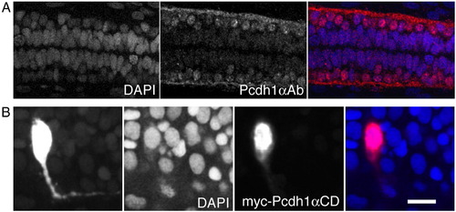
Pcdh1α intracellular domain is present in the nucleus. (A) Shown are dorsal views of the spinal cord from 24 hpf embryos; images are maximum intensity projections of two adjacent optical sections (1 μm spacing). The left panel shows DAPI labeling of cells. The middle panel shows the same field stained with antibody against Pcdh1αCD. The right panel is an overlay showing that Pcdh1α immunoreactivity is present in neuronal nuclei. (B) The leftmost panel is a maximum intensity projection of a spinal interneuron that has been labeled with a myc-tagged Pcdh1αCD. The myc-Pcdh1αCD fusion is distributed throughout the neuron. The middle two panels show single optical sections through the same neuron. The center-left panel shows staining of nuclei with DAPI and the center-right panel shows anti-myc immunolabeling. The myc-Pcdh1αCD is enriched in the nucleus. The overlay of the two fluorescence images is shown in the panel on the right. Scale bar = 10 μm. |
| Antibody: | |
|---|---|
| Fish: | |
| Anatomical Terms: | |
| Stage: | Prim-5 |
Reprinted from Developmental Biology, 321(1), Emond, M.R., and Jontes, J.D., Inhibition of protocadherin-alpha function results in neuronal death in the developing zebrafish, 175-187, Copyright (2008) with permission from Elsevier. Full text @ Dev. Biol.