
Fig. 4
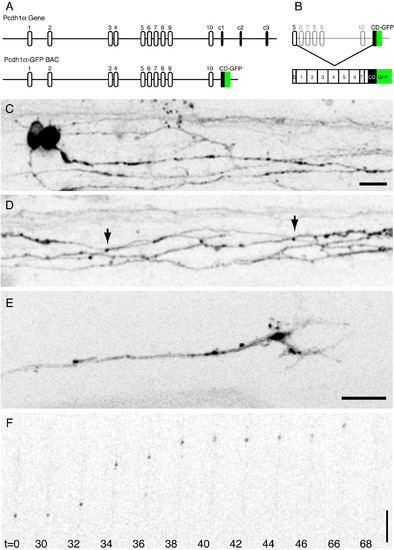
Pcdh1α-GFP expression by BAC recombineering. (A) The organization of the pcdh1α gene in the original (top) and recombineered (bottom) BACs. (B) Each variable exon consists of a signal sequence (S), 6 cadherin repeats, a transmembrane segment (T) and a very short portion of the cytoplasmic domain (CD). Each of these is spliced to the Pcdh1αCD-GFP to generate GFP-tagged proteins that are full length protocadherins. (C) Neurons labeled in the spinal cord of a ∼ 28 hpf embryo. Pcdh1α-GFP exhibits both nuclear and perinuclear staining and strongly labels axons. Scale bar = 10 μm. (D) Axonal labeling is largely diffuse and continuous, although small puncta are occasionally present (arrows). (E) Pcdh1α-GFP is enriched in the central core of axonal growth cones, and is likely concentrated in intracellular organelles. Scale bar = 8 μm. (F) Mobile transport organelles can be visualized using time-lapse microscopy. These transport packets have similar properties to transport packets previously observed both in vitro and in vivo. Scale bar = 10 μm. |
Reprinted from Developmental Biology, 321(1), Emond, M.R., and Jontes, J.D., Inhibition of protocadherin-alpha function results in neuronal death in the developing zebrafish, 175-187, Copyright (2008) with permission from Elsevier. Full text @ Dev. Biol.