
Fig. 1
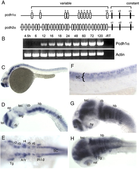
Pcdh1α is expressed in the early zebrafish nervous system. (A) Genomic organization of the two pcdhα genes. Most of this study is concerned with the pcdh1α gene. (B) RT-PCR with primers directed against the common cytoplasmic domain reveals the time-course of pcdh1α expression during development. Onset of expression occurs at ∼ 12 hpf, increases steadily for the first 3 days, then remains highly expressed. (C) Shown here is a 24 hpf embryo labeled with riboprobe against the constant cytoplasmic domain of the pcdh1α gene. Expression is confined to the developing nervous system. (D) Shown here is a lateral view of a brain dissected from a 24 hpf embryo. In the forebrain, expression is present in both the telencephalon and diencephalon. Expression is also found in the ventral midbrain, and in segmental clusters of cells in the hindbrain. In addition, pcdh1α is expressed in ephiphysial neurons. (E) In a dorsal view of the hindbrain, rhombomeric expression is revealed by ladder-like clusters of neurons. In addition, peripheral neurons also exhibit expression, including the trigeminal ganglion neurons, acoustico-vestibular neurons and neurons of the posterior lateral line ganglion. (F) Riboprobe against pcdh1α labels neurons throughout the developing spinal cord. (G and H) By 48 hpf, pcdh1α expression has expanded and has become very strong throughout the developing brain, including the tectum and cerebellum. In addition, staining can be seen in the retina. A/V, acoustico-vestibular; cb, cerebellum; di, diencephalon; ep, epiphysis; hb, hindbrain; hy, hypothalamus; pLLg, posterior lateral line ganglion; r2-r6, rhomobomeres 2–6; ret, retina; SC, spinal cord; tec, tectum; teg, tegmentum; tel, telencephalon; Tg, trigeminal ganglion. |
Reprinted from Developmental Biology, 321(1), Emond, M.R., and Jontes, J.D., Inhibition of protocadherin-alpha function results in neuronal death in the developing zebrafish, 175-187, Copyright (2008) with permission from Elsevier. Full text @ Dev. Biol.