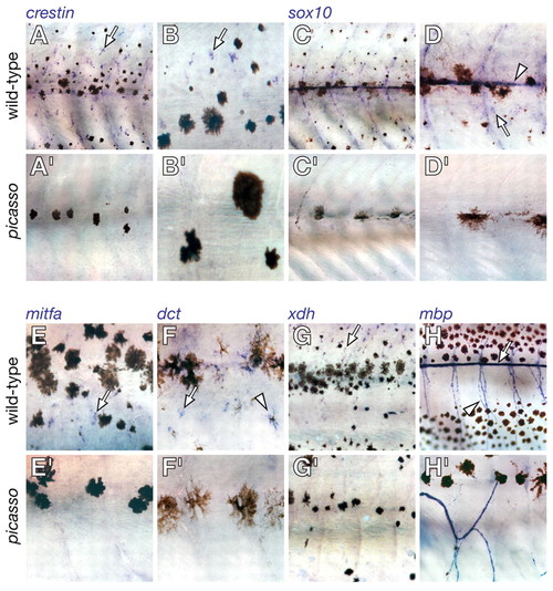
Metamorphic deficiencies for early and late markers of neural crest-derived lineages in picasso mutant larvae. Shown are corresponding regions of the mid-trunk for wild type (above) and picasso mutants during mid-metamorphosis (∼18 dpf). Individual melanophores are more spread in picasso mutants, typical of reduced melanophore densities (Parichy and Turner, 2003b; Parichy et al., 2003). (A,A′) crestin marks neural crest-derived cells in embryos (Luo et al., 2001) and identifies dispersed cells in the hypodermis of metamorphosing larvae (arrow). crestin+ cells were dramatically fewer in picasso mutants. (B,B′) Higher magnification image of crestin+ cells in wild type, and their absence in picasso. (C,C′) sox10 marks non-ectomesenchymal neural crest-derived cells in embryos, including pigment cell and glial precursors (Dutton et al., 2001; Gilmour et al., 2002), and identifies comparable populations in metamorphosing larvae (Parichy et al., 2003). sox10+ cells were fewer in picasso compared with wild type. (D,D′) Higher magnification showing sox10+ cells along the lateral line (arrowhead) and in the hypodermis (arrow) of wild type, but not picasso. (E,E′) mitfa marks melanophore and xanthophore precursors in embryos and is essential for melanoblast specification (Lister et al., 1999; Parichy et al., 2000b). mitfa+ cells (arrow) were numerous in wild type but not in picasso. (F,F′) dct identifies melanophore precursors (arrow) and melanophores (arrowhead) (Kelsh et al., 2000); dct+ cells were reduced or absent in picasso. (G,G′) Xanthine dehydrogenase (xdh) encodes an enzyme in the pteridine synthesis pathway of xanthophores (Parichy et al., 2000b). xdh+ cells (arrow) were numerous in wild type but transiently fewer in picasso. (H,H′) Myelin basic protein (mbp) marks mature glia in the peripheral nervous system (Brosamle and Halpern, 2002; Lyons et al., 2005) and mbp+ cells line the midbody lateral line (arrow) as well as ascending and descending nerve fibers (arrowhead). Although mbp+ cells were present in picasso, they were fewer in number and their associated nerves were misrouted.
|
