
FIGURE
Fig. 1
- ID
- ZDB-FIG-080711-9
- Publication
- Kimmel et al., 1979 - Target recognition in neurogenesis: formation of the Mauthner axon cap
- Other Figures
- All Figure Page
- Back to All Figure Page
Fig. 1
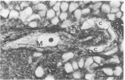
Axon cap in the zebrafish larva at 6 days after fertilization and left M cell (M) in transverse section. The M axon (arrow) enters the axon cap (ac) which is surrounded by glial cap cells (c). Dorsal is to the top; midline of the brain is immediately to the right of the figure. (Scale, 10 μm; 2.5-μm section ;phase-contrast with x100 oil immersion objective.) |
Expression Data
Expression Detail
Antibody Labeling
Phenotype Data
Phenotype Detail
Acknowledgments
This image is the copyrighted work of the attributed author or publisher, and
ZFIN has permission only to display this image to its users.
Additional permissions should be obtained from the applicable author or publisher of the image.
Full text @ Proc. Natl. Acad. Sci. USA