
FIGURE
Fig. 3
- ID
- ZDB-FIG-080711-11
- Publication
- Kimmel et al., 1979 - Target recognition in neurogenesis: formation of the Mauthner axon cap
- Other Figures
- All Figure Page
- Back to All Figure Page
Fig. 3
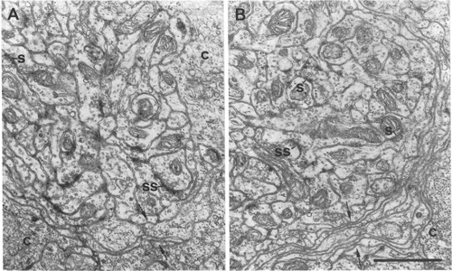
Electron micrographs of margins of normal (A) and M-cell deleted (B) axon caps. The thin sections were made after the caps shown in Figs. 1 and 2 (right side, M-cell deleted) were reembedded. Axo-axonal chemical synapses (s), including symmetrical synapses (ss), are present. Cytoplasmic extensions of the cap cells (c) form a lamellar structure surrounding the cap neuropil (arrows). The structure of the neuropil closely corresponds to the "core" portion of the axon cap of the adult goldfish described by Kohno (14); a "periphery" portion is absent in the zebrafish larva. (Scale, 1 μm.) |
Expression Data
Expression Detail
Antibody Labeling
Phenotype Data
Phenotype Detail
Acknowledgments
This image is the copyrighted work of the attributed author or publisher, and
ZFIN has permission only to display this image to its users.
Additional permissions should be obtained from the applicable author or publisher of the image.
Full text @ Proc. Natl. Acad. Sci. USA