
FIGURE
Fig. 4
- ID
- ZDB-FIG-080711-12
- Publication
- Kimmel et al., 1979 - Target recognition in neurogenesis: formation of the Mauthner axon cap
- Other Figures
- All Figure Page
- Back to All Figure Page
Fig. 4
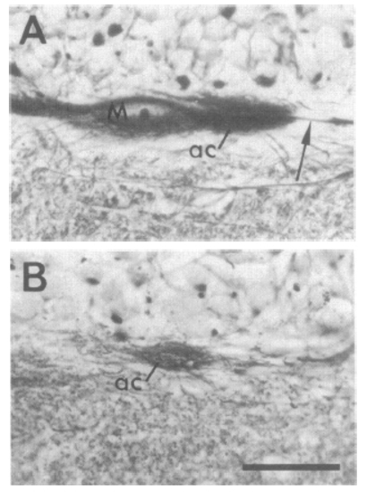
Light micrographs of M-axon caps in the axolotl larva. Both show transverse sections of the left side of the brain at the M-cell level (lesions made at stage 35); dorsal is to the top. (A) M cell (M) survived the lesion, and its axon (arrow) passes through the axon cap (ac) normally. (B) M cell was deleted. The cap neuropil (ac) is present at the correct position; it stains less densely than does the normal axon cap. (Scale, 50 μm; Rowell silver stain; bright-field illumination with X40 objective.) |
Expression Data
Expression Detail
Antibody Labeling
Phenotype Data
Phenotype Detail
Acknowledgments
This image is the copyrighted work of the attributed author or publisher, and
ZFIN has permission only to display this image to its users.
Additional permissions should be obtained from the applicable author or publisher of the image.
Full text @ Proc. Natl. Acad. Sci. USA