
- Title
-
Target recognition in neurogenesis: formation of the Mauthner axon cap
- Authors
- Kimmel, C.B., Sessions, S.K., and Kimmel, R.J.
- Source
- Full text @ Proc. Natl. Acad. Sci. USA
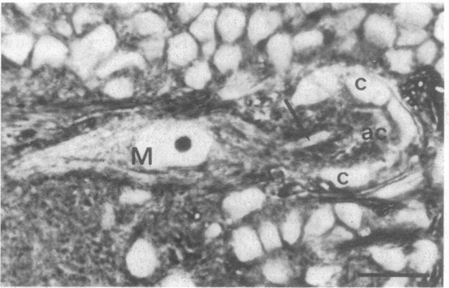
Axon cap in the zebrafish larva at 6 days after fertilization and left M cell (M) in transverse section. The M axon (arrow) enters the axon cap (ac) which is surrounded by glial cap cells (c). Dorsal is to the top; midline of the brain is immediately to the right of the figure. (Scale, 10 μm; 2.5-μm section ;phase-contrast with x100 oil immersion objective.) |
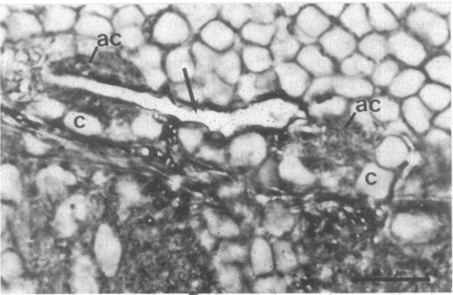
Left and right axon caps in a day 9 zebrafish larva in which the right M cell was deleted by microsurgery. The left M axon (arrow) passes through the left axon cap (ac), acquires a phase dark myelin sheath, and passes to the brain midline, where it leaves the plane of section (it does not enter the right axon cap). Both axon caps are surrounded by cap cells (c) and are similar in structure. Otherwise as in Fig. 1. (Scale, 10 μm.) |
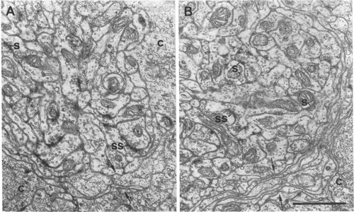
Electron micrographs of margins of normal (A) and M-cell deleted (B) axon caps. The thin sections were made after the caps shown in Figs. 1 and 2 (right side, M-cell deleted) were reembedded. Axo-axonal chemical synapses (s), including symmetrical synapses (ss), are present. Cytoplasmic extensions of the cap cells (c) form a lamellar structure surrounding the cap neuropil (arrows). The structure of the neuropil closely corresponds to the "core" portion of the axon cap of the adult goldfish described by Kohno (14); a "periphery" portion is absent in the zebrafish larva. (Scale, 1 μm.) |
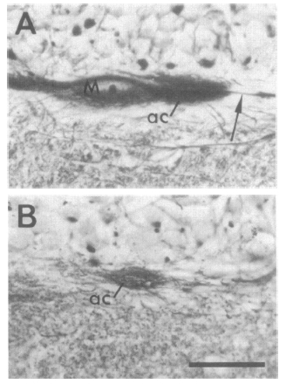
Light micrographs of M-axon caps in the axolotl larva. Both show transverse sections of the left side of the brain at the M-cell level (lesions made at stage 35); dorsal is to the top. (A) M cell (M) survived the lesion, and its axon (arrow) passes through the axon cap (ac) normally. (B) M cell was deleted. The cap neuropil (ac) is present at the correct position; it stains less densely than does the normal axon cap. (Scale, 50 μm; Rowell silver stain; bright-field illumination with X40 objective.) |