
FIGURE
Fig. 2
- ID
- ZDB-FIG-080711-10
- Publication
- Kimmel et al., 1979 - Target recognition in neurogenesis: formation of the Mauthner axon cap
- Other Figures
- All Figure Page
- Back to All Figure Page
Fig. 2
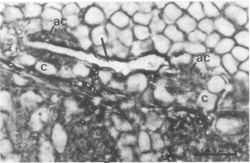
Left and right axon caps in a day 9 zebrafish larva in which the right M cell was deleted by microsurgery. The left M axon (arrow) passes through the left axon cap (ac), acquires a phase dark myelin sheath, and passes to the brain midline, where it leaves the plane of section (it does not enter the right axon cap). Both axon caps are surrounded by cap cells (c) and are similar in structure. Otherwise as in Fig. 1. (Scale, 10 μm.) |
Expression Data
Expression Detail
Antibody Labeling
Phenotype Data
Phenotype Detail
Acknowledgments
This image is the copyrighted work of the attributed author or publisher, and
ZFIN has permission only to display this image to its users.
Additional permissions should be obtained from the applicable author or publisher of the image.
Full text @ Proc. Natl. Acad. Sci. USA