|
Expression of NR Genes in Retina at 72 hpf
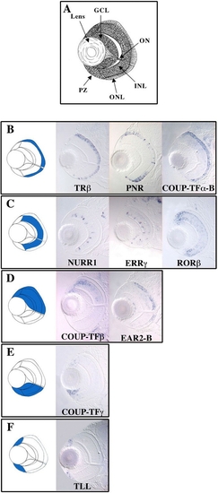
(A) Schematic of a zebrafish eye at 72 hpf showing the characteristic multilayered structure. GCL, ganglion cell layer; INL, inner nuclear layer; ONL, outer nuclear layer; ON, optic nerve; PZ, proliferative zone.
(B–F; left panels) Schematic showing in blue the various types of expression patterns found for NR genes in retinas at 72 hpf after whole-mount in situ hybridization and section. (B) Genes expressed in the ONL. (C) Genes expressed in the INL. (D) Genes expressed in the dorsal part of the retina. (E) Genes showing expression in the ventral part of the retina. (F) Genes showing expression in the proliferative zone of the retina.
|