|
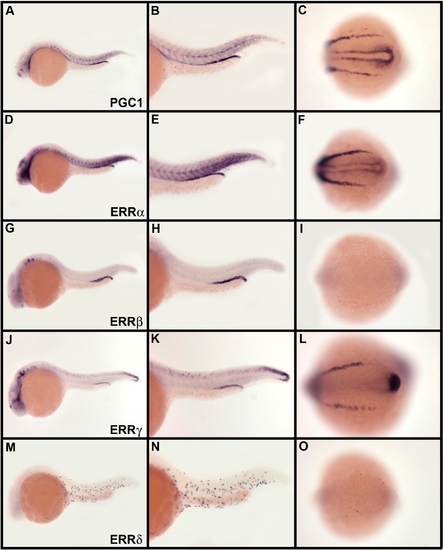
Overlapping Domains of Expression between PGC1 and ERRs
(A–O) Expression of PGC1 in slow muscle fibers, posterior pronephric ducts, mucous cells, epiphysis, and part of the telencephalon and diencephalon (A–C) overlaps extensively with the expression of ERRs. ERRα is coexpressed with PGC1 in slow muscle fibers, posterior pronephric ducts, telencephalon, and mucous cells (D–F). ERRβ is coexpressed with PGC1 in epiphysis and posterior pronephric ducts (G–I), ERRγ in epiphysis, part of the tegmentum, and posterior pronephric ducts and ERRδ in mucous cells. Embryos are at 24 hpf in lateral view anterior on the left except for (C, F, I, L, and O), which are shown at the 14-somite stage. Posterior part of the embryo is presented in dorsal view, anterior to the left. More extensive anatomical descriptions of these expression patterns are presented in Figure S3 and anatomical details are available at ZFIN (http://zfin.org)
|