
FIGURE
Fig. S10
- ID
- ZDB-FIG-080325-128
- Publication
- Lachnit et al., 2008 - Alterations of the cytoskeleton in all three embryonic lineages contribute to the epiboly defect of Pou5f1/Oct4 deficient MZspg zebrafish embryos
- Other Figures
- All Figure Page
- Back to All Figure Page
Fig. S10
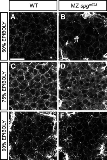
DEL cell morphology. Animal pole view confocal z-projections of Alexa488-Phalloidin stained embryos during epiboly. (A–F) Confocal sections of the DEL. A, C, and E show WT DEL cells. B, D, and F show MZspgm793 DEL cells. (A, B) At 60% epiboly; (C, D) at 75% epiboly; (E, F) at 90% epiboly. DEL morphology appears normal in MZspgm793 embryos. Scale bar: 20 μm. |
Expression Data
Expression Detail
Antibody Labeling
Phenotype Data
| Fish: | |
|---|---|
| Observed In: | |
| Stage Range: | Shield to 90%-epiboly |
Phenotype Detail
Acknowledgments
This image is the copyrighted work of the attributed author or publisher, and
ZFIN has permission only to display this image to its users.
Additional permissions should be obtained from the applicable author or publisher of the image.
Reprinted from Developmental Biology, 315(1), Lachnit, M., Kur, E., and Driever, W., Alterations of the cytoskeleton in all three embryonic lineages contribute to the epiboly defect of Pou5f1/Oct4 deficient MZspg zebrafish embryos, 1-17, Copyright (2008) with permission from Elsevier. Full text @ Dev. Biol.