
FIGURE
Fig. S7
- ID
- ZDB-FIG-080325-125
- Publication
- Lachnit et al., 2008 - Alterations of the cytoskeleton in all three embryonic lineages contribute to the epiboly defect of Pou5f1/Oct4 deficient MZspg zebrafish embryos
- Other Figures
- All Figure Page
- Back to All Figure Page
Fig. S7
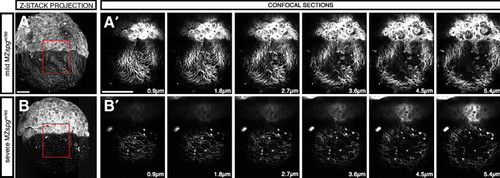
Excessive formation of cortical layer spherical structures. (A, B) Z-projection of anti-β-Tubulin immunofluorescence stain (Alexa488 conjugated α mouse-Ab) of MZspgm793 embryos. (A′, B′) Confocal slices depicting sphere localization within the cortical layer. (A) Mild MZspgm793 phenotype. (B) Severe MZspgm793 phenotype. Scale bar: 100 μm. |
Expression Data
| Antibody: | |
|---|---|
| Fish: | |
| Anatomical Terms: | |
| Stage: | Shield |
Expression Detail
Antibody Labeling
Phenotype Data
| Fish: | |
|---|---|
| Observed In: | |
| Stage: | Shield |
Phenotype Detail
Acknowledgments
This image is the copyrighted work of the attributed author or publisher, and
ZFIN has permission only to display this image to its users.
Additional permissions should be obtained from the applicable author or publisher of the image.
Reprinted from Developmental Biology, 315(1), Lachnit, M., Kur, E., and Driever, W., Alterations of the cytoskeleton in all three embryonic lineages contribute to the epiboly defect of Pou5f1/Oct4 deficient MZspg zebrafish embryos, 1-17, Copyright (2008) with permission from Elsevier. Full text @ Dev. Biol.