
FIGURE
Fig. S2
- ID
- ZDB-FIG-080325-115
- Publication
- Lachnit et al., 2008 - Alterations of the cytoskeleton in all three embryonic lineages contribute to the epiboly defect of Pou5f1/Oct4 deficient MZspg zebrafish embryos
- Other Figures
- All Figure Page
- Back to All Figure Page
Fig. S2
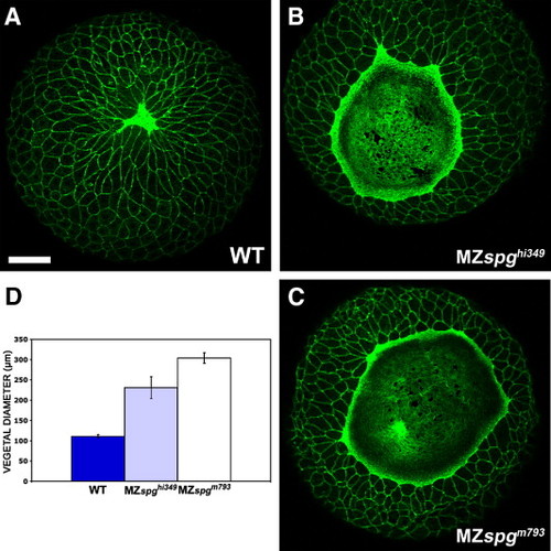
Comparison of delay in EVL epiboly in MZspghi349 and MZspgm793 embryos. Embryos were stained with Alexa488-Phalloidin and z-projections of confocal z-stacks generated. (A-C) Vegetal view of 95% epiboly stage embryos. (A) WT; (B) MZspghi349; (C) MZspgm793. Scale bar in A (for A-C): 100 μm. (D) Measurement of the distance between opposing EVL margins including SE. WT = 111 ± 4.2 μm; MZspghi349 = 231 ± 27 ¼m; MZspgm793 = 304 ± 13 µm. Scale bar: 20 μm. |
Expression Data
Expression Detail
Antibody Labeling
Phenotype Data
| Fish: | |
|---|---|
| Observed In: | |
| Stage: | 90%-epiboly |
Phenotype Detail
Acknowledgments
This image is the copyrighted work of the attributed author or publisher, and
ZFIN has permission only to display this image to its users.
Additional permissions should be obtained from the applicable author or publisher of the image.
Reprinted from Developmental Biology, 315(1), Lachnit, M., Kur, E., and Driever, W., Alterations of the cytoskeleton in all three embryonic lineages contribute to the epiboly defect of Pou5f1/Oct4 deficient MZspg zebrafish embryos, 1-17, Copyright (2008) with permission from Elsevier. Full text @ Dev. Biol.