
FIGURE
Fig. S3
- ID
- ZDB-FIG-080325-117
- Publication
- Lachnit et al., 2008 - Alterations of the cytoskeleton in all three embryonic lineages contribute to the epiboly defect of Pou5f1/Oct4 deficient MZspg zebrafish embryos
- Other Figures
- All Figure Page
- Back to All Figure Page
Fig. S3
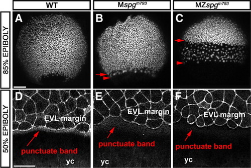
Maternal Pou5f1 impact on the DEL and EVL phenotype. (A–C) Z-projection of confocal image stacks of 85% epiboly embryos stained with Sytox green to visualize nuclei. (A) WT; (B) Mspgm793; (C) MZspgm793. A delay of EVL and DEL is detectable based on altered distribution of nuclei. (D–F) Higher magnification of the EVL margin of confocal z-projections of Alexa488-Phallodin stained embryos. (D) WT; (E) Mspgm793; (F) MZ spgm793. Note delay in change of marginal EVL cell shape and reduced stain intensity of the punctate F-Actin band in the YSL. yc: yolk cell. Scale bar: 100 μm. |
Expression Data
Expression Detail
Antibody Labeling
Phenotype Data
| Fish: | |
|---|---|
| Observed In: | |
| Stage Range: | 50%-epiboly to 75%-epiboly |
Phenotype Detail
Acknowledgments
This image is the copyrighted work of the attributed author or publisher, and
ZFIN has permission only to display this image to its users.
Additional permissions should be obtained from the applicable author or publisher of the image.
Reprinted from Developmental Biology, 315(1), Lachnit, M., Kur, E., and Driever, W., Alterations of the cytoskeleton in all three embryonic lineages contribute to the epiboly defect of Pou5f1/Oct4 deficient MZspg zebrafish embryos, 1-17, Copyright (2008) with permission from Elsevier. Full text @ Dev. Biol.