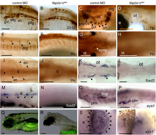
Neural crest and placode derivatives are absent or reduced in tfap2a/c deficient embryos. (A, B) Lateral views of 28 hpf embryos processed for anti-HNK1 (Zn12 antibody) immunoreactivity (IR), revealing the presence of cranial ganglia neurons in panel A, a control MO-injected wild-type embryo, and their absence in panel B, a tfap2a-/cMO embryo (9 of 10 tfap2a-/cMO embryos). (C–F) Lateral and (G–J) ventral views of 72 hpf embryos processed for anti-Hu IR. (C, E, G, I) In control embryos anti-Hu IR reveals panel C, cranial ganglia surrounding the otic vesicle (ot), (E), dorsal root ganglia (drg) in the trunk, (G), sympathetic neurons at vagal level, and (I), proximal enteric neurons, adjacent to gut (g), also at vagal level. (D, F, H, J) All of these neurons appear to be absent from a tfap2a-/cMO embryo (8 of 8 tfap2a-/cMO embryos). (K, L) Dorsal and (M, N) lateral views of 28 hpf embryos processed to reveal foxd3 expression, which labels (K) satellite cells (arrowheads) and (M) Schwann cells associated with lateral line processes (arrowheads) in a control embryo, but not in panels L, N, a tfap2a-/cMO embryo (12 of 12 tfap2a-/cMO embryos). (O, P) Lateral views of 28 hpf embryos processed to reveal eya1 expression, which is detected in the lateral line migrating primordium (llmp) in panel O, a control embryo, but absent in panel P, a tfap2a-/cMO embryo (11 of 11 tfap2a-/cMO MO embryos). eya1 expression in presumed pectoral fin mesoderm (pfm) remains normal in the tfap2a-/cMO MO embryo (11 of 11 tfap2a-/cMO MO embryos). (Q, R) Lateral head views of 4 dpf embryos incubated in DASPEI, which is taken up by hair cells. In panel Q, a control embryo, DASPEI-positive hair cells in the inner ear (arrowhead) and the neuromasts (arrows) are visible. In panel R, a tfap2a-/cMO embryo, DASPEI positive cells are not seen. (S, T) Dorsal anterior views of 28 hpf embryos processed to reveal eya1 expression. eya1 expression in olfactory placode is apparent in panel S, a control embryo. In panel R, a tfap2a-/cMO embryo, a small cluster of eya1-expressing cells is present in this position (11 of 11 tfap2a-/cMO embryos). gV, trigeminal ganglion; allg, anterior lateral line ganglion; ac, acoustic ganglion; pllg, posterior lateral line ganglion; gAD, anterodorsal lateral line ganglion; gAV, anteroventral lateral line ganglion; gVII, facial sensory ganglion; gIX, glossopharyngeal ganglion; gX, vagus ganglion; e, eye; ot, otic vesicle. Scale bars: (A–R), 100 μm; (S, T), 50 μm.
|
