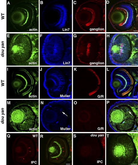
Retinal cell specification is delayed by the dou yan mutation. A-D: At 78 hours postfertilization (hpf) in wild-type retinas, Lin7 expression (blue) is concentrated in the bipolar cell body region and the plexiform layers. The ganglion cells stained with zn8 antibody (red) localize to the basal region of the retinas. The actin distribution as revealed by phalloidin staining highlights the plexiform layers. E-H: In dou yan mutant retina at 78 hpf, Lin7 (blue) is expressed in the entire retina with increased accumulation in the basal retinal region. This expression pattern is similar to Lin7 expression in wild-type retinas at 48 hpf (Wei et al.,[2006a]). A patch of retinal cells that express zn8 antigen is noticeable at the basal region of the retina in the mutants. I-L: Muller cells (blue) and green/red double cones (G/R, red) are properly differentiated in the wild-type retinas at 78 hpf. M-P: In most of the mutant retinas at 78 hpf, only a few cells display positive expression of Muller cell marker carbonic anhydrase (arrows, blue) and the double cone marker Zpr1 was not detectable. Q-T: Interplexiform layer cells (IPC, red) are present in wild-type retinas (Q,R) but not in the mutant retina (S,T) at 78 hpf. Scale bars = 20 μm.
|
