|
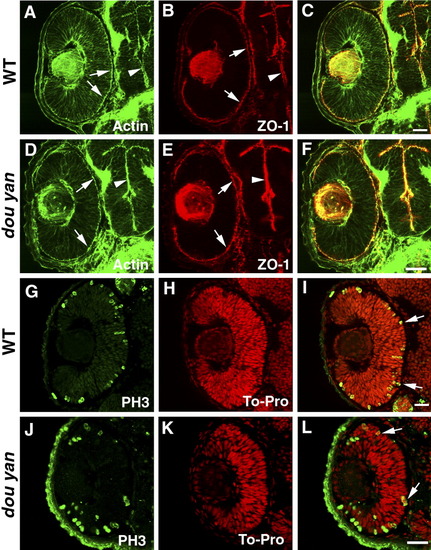
The dou yanmi234 mutation does not affect apical epithelial polarity. A-C: Apical localization of adherens junctions in the retina (arrows) and brain (arrowheads) of the 48 hours postfertilization (hpf) wild-type embryos is visualized by the staining patterns of ZO-1 (red) and adherens junction-associated actin bundles (A, green, arrows). D-F: ZO-1 (red) and adherens junction-associated actin bundles (green) localize properly to the apical surface of the retina (arrows) and brain (arrowheads) in dou yanmi234 embryos at 48 hpf. G-L: In 48 hpf retinas, the M-phase nuclei as visualized with anti-phosphorylated-Histone H3 antibody (PH3, green) localize to the apical regions of the retinas of mutant (J-L) and wild-type (G-I) embryos. Cell nuclei were labeled with TO-PRO (red). Scale bars = 20 μm.
|