FIGURE SUMMARY
Title

Vascular defects of DYRK1A knockouts are ameliorated by modulating calcium signaling in zebrafish

Authors
Cho, H.J., Lee, J.G., Kim, J.H., Kim, S.Y., Huh, Y.H., Kim, H.J., Lee, K.S., Yu, K., Lee, J.S.
Source
Full text @ Dis. Model. Mech.

dyrk1aakrb1 mutant embryos show cerebral hemorrhagic phenotype and abnormal development of CtAs in the brain. (A) Cerebral hemorrhage was observed in dyrk1aakrb1 mutant embryos (dyrk1aakrb1) at 52 hpf (Ac-Af, arrows) compared to WT (Aa, Ab). Aa,Ac,Ae show the lateral view; Ab,Ad,Af show the dorsal view. (B) Embryonic cerebral hemorrhage of WT occurred spontaneously in 9.9% of embryos, whereas dyrk1aakrb1 embryos showed cerebral hemorrhage of 27.9% penetrance (Normal), using o-dianisidine staining. The cerebral hemorrhage of WT and dyrk1aakrb1 mutants increased up to 16.3% and 40.5%, respectively, by inducing heat stress with 2.5 h incubation at 35°C from 48-50.5 hpf (Heat-stressed). The mean percentages for each genotype were presented from four independent experiments with approximately 20 embryos for each repeat. (C) Confocal fluorescent images at 52 hpf showing the development of CtAs in WT and dyrk1aakrb1 mutant embryos in the Tg(kdrl:EGFP) background. (D) The lengths and branching points of CtAs in dyrk1aakrb1 mutants were reduced down to 70.2% and 73.3%, respectively, compared to the WT embryos as 100% at 52 hpf. *P<0.05, ***P<0.005 (Mann–Whitney U test). Data are mean±s.e.m. fb, forebrain; mb, midbrain; hb, hindbrain; e, eye; y, yolk. Scale bar: 250 µm in A; 50 µm in C.

Zebrafish dyrk1aa and dyrk1ab are expressed in the developing brain during embryogenesis and detected in endothelial cells of the vasculature. (A) WISH showed that dyrk1aa was expressed in the forebrain (black arrowheads, Aa-Af; black brackets, Ag-Al), the midbrain (gray arrowheads, Aa-Af; gray brackets, Ag-Al), the hindbrain (blue arrowheads, Aa-Af; blue brackets, Ag-Al) at 24, 48 and 72 hpf, and the spinal cord (orange arrowheads, Aa and Ab) at 24 hpf. It was also detected in the heart (black asterisks, Ad,Af,Aj,Al) and in the retina (red arrows, Ai-Al) at 48 and 72 hpf. Am and An are sectioned images of WISH embryos showing the expression of dyrk1aa in the tectum (green arrows), tegmentum (black arrow), the ganglionic cell layer (purple arrows) and the inner nuclear layer of the retina (red arrows) at 48 hpf and 72 hpf. See Fig. S3 for WISH of dyrk1ab. (B) Semi-quantitative RT-PCR analysis of dyrk1aa and dyrk1ab mRNA expression in whole embryos at indicated developmental stages. dyrk1aa and dyrk1ab expression was observed at the one-cell stage followed by diminishing at 6 hpf and resuming after 24 hpf. (C) Semi-quantitative RT-PCR analysis using endothelial cells isolated by FACS for sorting GFP-positive and -negative cells of Tg(kdrl:EGFP) embryos at 48 hpf revealed that dyrk1aa and dyrk1ab mRNAs were expressed in GFP-positive endothelial cells as well as in GFP-negative embryonic cells. Scale bars: 200 µm in Aa-Al; 50 µm in Am,An.

The cerebral hemorrhage and CtA angiogenic defects in dyrk1aakrb1 mutants can be rescued by dyrk1aa expression, with the kinase activity of Dyrk1aa required for its phenotypic rescues. (A) The o-dianisidine staining images at 52 hpf showed that the cerebral hemorrhage (arrows) of dyrk1aakrb1 embryos was rescued by WT dyrk1aa mRNA expression, but not in no-injection control, K193R-dyrk1aa or mCherryRed mRNA. (B) The frequency of cerebral hemorrhage of dyrk1aakrb1 was reduced from 41.7% to 25.4% by injecting WT dyrk1aa mRNA of 0.1 ng, but not significantly changed by injecting K193R-dyrk1aa or mCherryRed mRNA at 52 hpf. (C) Injection of mRNAs of WT dyrk1aa, K193R-dyrk1aa or mCherryRed control did not affect the cerebral hemorrhage in WT embryos at 52 hpf. The mean percentages for each genotype were presented from three independent experiments with approximately 20 embryos in each repeat. (D) The compiled images of CtAs of Tg(kdrl:EGFP) at 52 hpf by confocal microscopy showed that the angiogenic defects of the CtAs of dyrk1aakrb1 embryos were rescued by WT dyrk1aa mRNA injection. (E) The mean percentages of length and branching points of CtAs in dyrk1aakrb1 mutants were rescued from 66.5% to 83.4% and from 61.2% to 108.3%, respectively, with 0.05 ng of dyrk1aa mRNA injection, relative to WT as 100%, and rescued to 89.9% and 122.0%, respectively, with 0.1 ng of dyrk1aa mRNA injection. Expression with 0.2 ng of dyrk1aa mRNA did not exhibit the rescue effect. Expression of dyrk1aa mRNA with the same doses in WT embryos also increased the length and branching points of CtAs in a dose-dependent manner (113.8% and 139.6% with 0.2 ng of dyrk1aa mRNA, respectively). *P<0.05, **P<0.01, ***P<0.005 (one-way ANOVA). n.s., not significant. Data are mean±s.e.m. Scale bar: 100 µm in A; 50 µm in D.

The transmission electron microscopy revealed that dyrk1aakrb1 embryos had abnormal vessel walls in the brain at 52 hpf. (A,B) Brain vessels in WT and dyrk1aakrb1. A′ and B′ show enlarged images of the boxed areas in A and B, respectively. (A′,B′) Arrows in A′ indicate the compact structure of vessel walls, whereas arrowheads in B′ designate the loose connection of vessel walls. Dashed lines demarcate the border between the vessel walls and the lumen. EC, endothelial cell; LM, vessel lumen; RBC, red blood cell; VW (dotted arrows), vessel wall. Scale bars: 1 µm.

PHENOTYPE:
Fish:
Observed In:
Stage: Long-pec

Inhibition of DYRK1A by harmine induces brain hemorrhage and vasculature defects in the hindbrain. (A) WT Tg(kdrl:EGFP) embryos were treated with increasing concentrations of DYRK1A inhibitor harmine, from 24 hpf until 52 hpf: Aa, DMSO; Ab, 10 µM harmine; Ac, 25 µM harmine; Ad, 50 µM harmine. Vascular patterning defects of CtAs are shown (red brackets) by confocal imaging at 52 hpf (lateral view). (B) Quantification of the effect on number of developing CtAs by harmine treatment. The numbers of CtAs were dramatically reduced by treating harmine in a dose-dependent manner. n=11 each group. (C) Quantification of the brain hemorrhage penetrance. The cerebral hemorrhagic phenotype of 2.3% in DMSO-treated embryos was increased from 14.8% to 65.7% by harmine treatment from 10 µM to 50 µM. The mean percentage for each treatment was shown from three independent experiments with approximately 40 embryos in each repeat. (D) Schematic showing the strategy of the in vivo chemical library screening to identify small molecule modifiers for cerebral hemorrhagic phenotype upon DYRK1A inhibition. Data are mean±s.e.m. Scale bar: 100 µm.

EXPRESSION / LABELING:
Gene:
Fish:
Condition:
Anatomical Term:
Stage: Long-pec
PHENOTYPE:
Fish:
Condition:
Observed In:
Stage: Long-pec

EGTA identified by the chemical library screening effectively rescues the cerebral hemorrhage and abnormal CtA development of dyrk1aakrb1 mutants. (A) o-dianisidine staining images at 52 hpf showing that the cerebral hemorrhage (arrow) of dyrk1aakrb1 embryos were rescued by treating with 10 nM EGTA. WT embryos were not affected by the treatment at the same concentration. (B) Quantitation of the rescue of the cerebral hemorrhagic phenotype by EGTA. The cerebral hemorrhage in dyrk1aakrb1 embryos is reduced from 37.5% to 23.6% by 10 nM EGTA treatment. The data is presented with five independent experiments with approximately 20 embryos in each repeat. (C) Compiled confocal microscopy images of the rescue effect on angiogenic defects of CtAs in dyrk1aakrb1 embryos by EGTA treatment. (D) Quantitative data showing that the reduced mean percentages of length and branching points of CtAs in dyrk1aakrb1 embryos at 52 hpf (76% and 57.4%, respectively, compared to WT embryos as 100%) were increased up to 86.6% and 85.9%, respectively, by 10 nM EGTA treatment. n≥11 each group. (E) Schematic depicts the treatment scheme of EGTA at developmental stages from 24 to 52 hpf in dyrk1aakrb1 embryos. (F) The cerebral hemorrhage of dyrk1aakrb1 embryos at 52 hpf (arrows) were rescued by 10 nM EGTA treatment for 24-32 hpf but not by the treatment for 32-48 hpf. (G) Quantitative data showing that cerebral hemorrhage of dyrk1aakrb1 embryos reduced from 37.7% to 26.3% by treating with 10 nM EGTA for 24-32 hpf. *P<0.05, **P<0.01, ***P<0.005 (one-way ANOVA). n.s., not significant. Data are mean±s.e.m. Scale bars: 100 µm in A,F; 50 µm in C.

FK506 rescues the cerebral hemorrhage and CtA angiogenic defects in dyrk1aakrb1 mutants. (A) The cerebral hemorrhage of dyrk1aakrb1 mutant embryos (arrows) was rescued by the treatment of 50 ng/ml FK506. (B) The cerebral hemorrhage in dyrk1aakrb1 mutant embryos at 52 hpf (40.5%) was reduced to 17.8% and 13.4% by the treatment of 50 ng/ml and 100 ng/ml FK506, respectively. No differences were seen in WT with the same treatments. (C) The compiled confocal microscopy images of CtAs in the Tg(kdrl:EGFP) background showed that CtA defects in dyrk1aakrb1 embryos were rescued by FK506 treatment in a dose-dependent manner, and CtA angiogenesis in WT was also affected. (D) The reduced mean percentage of CtA length of dyrk1aakrb1 mutants (75.5%) was significantly rescued up to 84.6% by 50 ng/ml FK506, whereas that of mutant branching points (74.3%) was significantly rescued up to 96.4% by 100 ng/ml FK506 at 52 hpf, compared to WT embryos as 100%. 50 ng/ml FK506 treatment increased the WT CtA length up to 110.3%, whereas 100 ng/ml FK506 treatment increased both length and branching points of WT CtAs (109.5% and 133.8%, respectively). *P<0.05, **P<0.01, ***P<0.005 (one-way ANOVA). n.s., not significant. Data are mean±s.e.m. Scale bars: 100 µm in A; 50 µm in C.

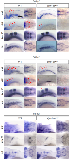
Expression of vascular markers is reduced in dyrk1aakrb1 mutant embryos, but the hindbrain development is not affected. The vascular markers of kdrl and dll4 were reduced at 30 and 36 hpf (red arrows), and kdrl expression was decreased in 52 hpf. The markers of krox20 (red asterisks) and isl1 were unchanged in dyrk1aakrb1 mutant embryos. Scale bars: 100 μm

dyrk1aa mutation does not affect the heart rates of zebrafish embryos at 52 hpf. (A) Heart rates were measured in WT and dyrk1aakrb1 embryos under a light microscope during 11 seconds at RT. N=7 each genetic group. (B) The captured images to measure the heart rates of WT and dyrk1aakrb1 embryos (refer to the Movies 1 and 2 for actual movies). p-values by Mann-Whitney U test: n.s., not significant. Data are mean ±s.e.m. Scale bars: 250 μm

PHENOTYPE:
Fish:
Observed In:
Stage: Long-pec

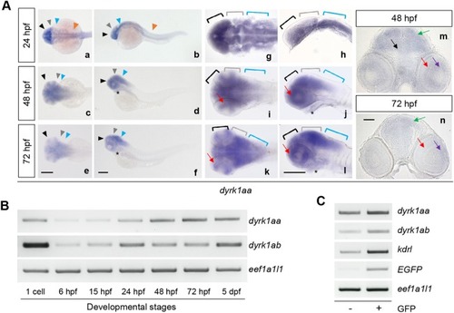
dyrk1ab is expressed in the developing brain region. By WISH (whole mount in situ hybridization), dyrk1ab was expressed in the forebrain (black arrowheads, a-f; black brackets, g-l), the midbrain (gray arrowheads, a-f; gray brackets, g-l), the hindbrain (blue arrowheads, a-f; blue brackets, g-l) at 24, 48 and 72 hpf and the spinal cord (orange arrowheads, a and b) at 24 hpf. It was also detected in the heart (asterisks, d, f, j and l) and in the retina (red arrows, i-l) at 48 and 72 hpf. (m and n) Sectioned images of WISH embryos showed the expression of dyrk1ab in the tectum (green arrows) and the retina (red arrows) at 48 hpf and 72 hpf. Scale bars: 200 μm in (a-l) and 50 μm in (m and n)

EXPRESSION / LABELING:
Gene:
Fish:
Anatomical Terms:
Stage Range: Prim-5 to Protruding-mouth

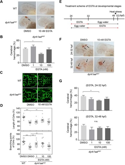
The vascular phenotype by EGTA treatment is not affected in WT embryos. No differences were observed in the mean percentages of cerebral hemorrhage in WT embryos (A) and the mean percentages of length and branching points of CtAs (C) by 1, 10 and 100 nM EGTA treatment. (B) The compiled images of CtAs by confocal microscopy show that the development of the CtAs with dyrk1aakrb1 embryos are rescued by 1 nM and 10 nM of EGTA treatment in the Tg(kdrl:EGFP) background (see Fig. 6D). WT embryos are not affected by EGTA treatment. p-values by one-way ANOVA: n.s., not significant. Data are mean±s.e.m. Scale bar: 50 μm.

ZFIN is incorporating published figure images and captions as part of an ongoing project. Figures from some publications have not yet been curated, or are not available for display because of copyright restrictions.

Acknowledgments
This image is the copyrighted work of the attributed author or publisher, and ZFIN has permission only to display this image to its users. Additional permissions should be obtained from the applicable author or publisher of the image. Full text @ Dis. Model. Mech.