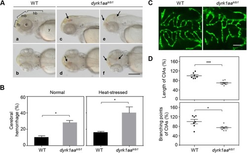
dyrk1aakrb1 mutant embryos show cerebral hemorrhagic phenotype and abnormal development of CtAs in the brain. (A) Cerebral hemorrhage was observed in dyrk1aakrb1 mutant embryos (dyrk1aakrb1) at 52 hpf (Ac-Af, arrows) compared to WT (Aa, Ab). Aa,Ac,Ae show the lateral view; Ab,Ad,Af show the dorsal view. (B) Embryonic cerebral hemorrhage of WT occurred spontaneously in 9.9% of embryos, whereas dyrk1aakrb1 embryos showed cerebral hemorrhage of 27.9% penetrance (Normal), using o-dianisidine staining. The cerebral hemorrhage of WT and dyrk1aakrb1 mutants increased up to 16.3% and 40.5%, respectively, by inducing heat stress with 2.5 h incubation at 35°C from 48-50.5 hpf (Heat-stressed). The mean percentages for each genotype were presented from four independent experiments with approximately 20 embryos for each repeat. (C) Confocal fluorescent images at 52 hpf showing the development of CtAs in WT and dyrk1aakrb1 mutant embryos in the Tg(kdrl:EGFP) background. (D) The lengths and branching points of CtAs in dyrk1aakrb1 mutants were reduced down to 70.2% and 73.3%, respectively, compared to the WT embryos as 100% at 52 hpf. *P<0.05, ***P<0.005 (Mann–Whitney U test). Data are mean±s.e.m. fb, forebrain; mb, midbrain; hb, hindbrain; e, eye; y, yolk. Scale bar: 250 µm in A; 50 µm in C.
|
