
|
Fig. S5
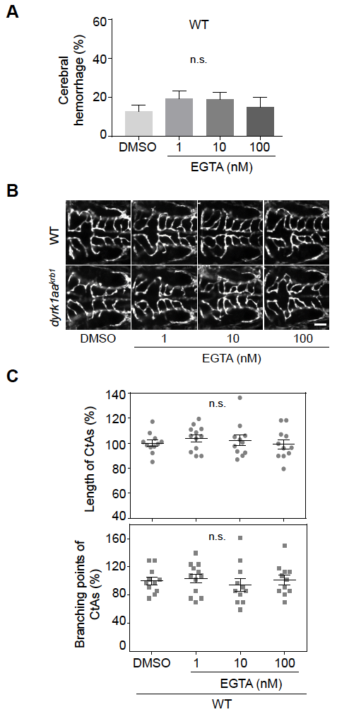
The vascular phenotype by EGTA treatment is not affected in WT embryos. No differences were observed in the mean percentages of cerebral hemorrhage in WT embryos (A) and the mean percentages of length and branching points of CtAs (C) by 1, 10 and 100 nM EGTA treatment. (B) The compiled images of CtAs by confocal microscopy show that the development of the CtAs with dyrk1aakrb1 embryos are rescued by 1 nM and 10 nM of EGTA treatment in the Tg(kdrl:EGFP) background (see Fig. 6D). WT embryos are not affected by EGTA treatment. p-values by one-way ANOVA: n.s., not significant. Data are mean±s.e.m. Scale bar: 50 μm.