
- Title
-
Ultrastructural Alterations in Thyrocytes of Zebrafish ( Danio rerio) after Exposure to Propylthiouracil and Perchlorate
- Authors
- Schmidt, F., Wolf, R., Baumann, L., Braunbeck, T.
- Source
- Full text @ Toxicol. Pathol.
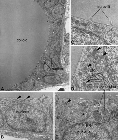
Ultrastructure of thyroidal tissue in control zebrafish (Danio rerio). The epithelium encloses an evenly stained colloid devoid of inclusions (A). The nucleus is basally located and most organelles can be found in apical position (B, E). Mitochondria appear spherically to ovally shaped; the rough endoplasmic reticulum and Golgi fields (*) are of cistern-like appearance (D, E). At the apical pole of thyrocytes, few electron-dense lysosomes are detectable (▸), and at the border to the colloid, some microvilli are detectable (B, C, D, E). Magnifications: A: 2,000×; B: 10,000×; C: 12,500×; D: 40,000×; E: 31,500×. |
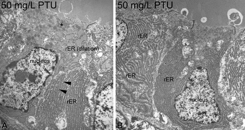
Ultrastructure of propylthiouracil-exposed zebrafish thyroids. At 50 mg/L, an electron-dense cytoplasm and shrunken nuclei present first symptoms of degeneration (A). Increased amounts of heterochromatin are visible (A, B). Marked proliferation, dilation, and fenestration in the rough endoplasmic reticulum (▸) are further alterations (A). The apical regions display proliferations of lysosomes (*; A). Magnifications: A: 10,000×; B: 4,000×. |
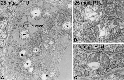
Mitochondrial alterations in zebrafish exposed to propylthiouracil. Already at 2.5 mg/L, mitochondria showed irregular swelling of the intercristae space (C). Higher exposure groups displayed proliferations and extensive swellings (*; A, B). Furthermore, dilation of the rough endoplasmic reticulum is visible (A). Magnifications: A: 10,000×; B and C: 40,000×. |
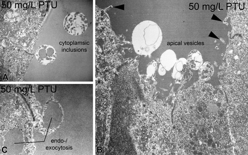
Apical alterations in zebrafish thyrocytes caused by propylthiouracil exposure. At concentrations ≥10 mg/L, cytoplasmic inclusions were evident (A). At concentrations ≥2.5 mg/L, numerous apical vesicles were seen protruding into the follicular lumen (B). Bleb-like structures indicate endo- or exocytotic processes at concentrations ≥10 mg/L (B, C). Moderate proliferation of microvilli can be observed at concentrations ≥10 mg/L (▸; B). Magnifications: A: 12,500×; B: 8,000×; C: 20,000×. |
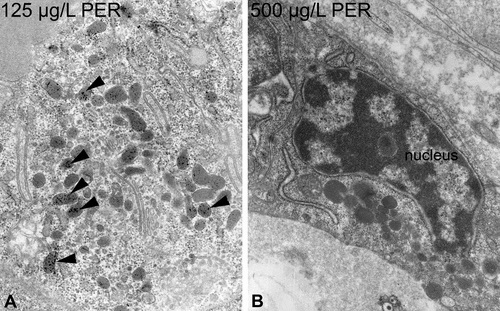
Further alterations of perchlorate-exposed zebrafish thyrocytes comprise a moderate increase in lipofuscin within lysosomes (▸; A). Moreover, the nucleus showed increased amounts of heterochromatin and an irregular outline, especially at 125 and 500 µg/L (B). Magnification: A: 20,000×; B: 12,500×. |
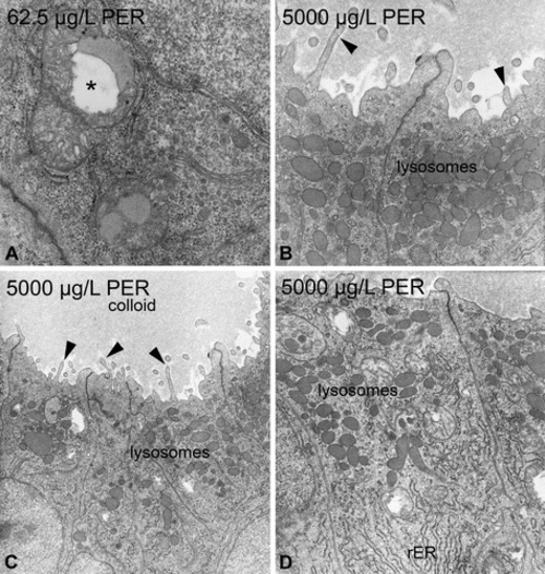
Perchlorate-induced ultrastructural alterations in zebrafish thyrocytes. At concentrations ≥62.5 µg/L, mitochondria are swollen and display irregular swellings of the intercristae space (*; A). At concentrations of 5,000 µg/L, a marked increase in lysosomes mostly located in the apical part of thyrocytes was visible (B, C, D). The rough endoplasmic reticulum showed moderate proliferation and some fenestration (D). The electron density of the colloid markedly decreased in higher concentration groups (C). Proliferations of microvilli are observable in concentrations ≥62.5 µg/L (▸; B, C). Magnification: A: 31,500×; B: 20,000×; C: 10,000×; D: 16,000×. |