
FIGURE
Fig. 3
- ID
- ZDB-FIG-170921-82
- Publication
- Schmidt et al., 2017 - Ultrastructural Alterations in Thyrocytes of Zebrafish ( Danio rerio) after Exposure to Propylthiouracil and Perchlorate
- Other Figures
- All Figure Page
- Back to All Figure Page
Fig. 3
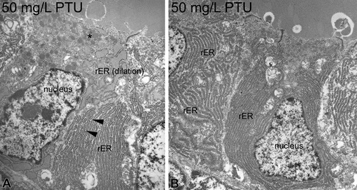
Ultrastructure of propylthiouracil-exposed zebrafish thyroids. At 50 mg/L, an electron-dense cytoplasm and shrunken nuclei present first symptoms of degeneration (A). Increased amounts of heterochromatin are visible (A, B). Marked proliferation, dilation, and fenestration in the rough endoplasmic reticulum (▸) are further alterations (A). The apical regions display proliferations of lysosomes (*; A). Magnifications: A: 10,000×; B: 4,000×. |
Expression Data
Expression Detail
Antibody Labeling
Phenotype Data
Phenotype Detail
Acknowledgments
This image is the copyrighted work of the attributed author or publisher, and
ZFIN has permission only to display this image to its users.
Additional permissions should be obtained from the applicable author or publisher of the image.
Full text @ Toxicol. Pathol.