
FIGURE
Fig. 1
- ID
- ZDB-FIG-170921-80
- Publication
- Schmidt et al., 2017 - Ultrastructural Alterations in Thyrocytes of Zebrafish ( Danio rerio) after Exposure to Propylthiouracil and Perchlorate
- Other Figures
- All Figure Page
- Back to All Figure Page
Fig. 1
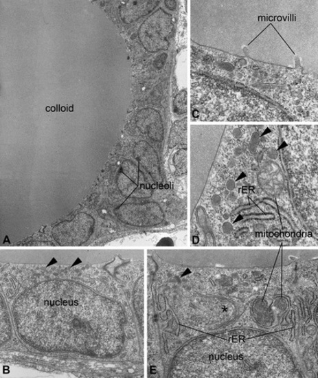
Ultrastructure of thyroidal tissue in control zebrafish (Danio rerio). The epithelium encloses an evenly stained colloid devoid of inclusions (A). The nucleus is basally located and most organelles can be found in apical position (B, E). Mitochondria appear spherically to ovally shaped; the rough endoplasmic reticulum and Golgi fields (*) are of cistern-like appearance (D, E). At the apical pole of thyrocytes, few electron-dense lysosomes are detectable (▸), and at the border to the colloid, some microvilli are detectable (B, C, D, E). Magnifications: A: 2,000×; B: 10,000×; C: 12,500×; D: 40,000×; E: 31,500×. |
Expression Data
Expression Detail
Antibody Labeling
Phenotype Data
Phenotype Detail
Acknowledgments
This image is the copyrighted work of the attributed author or publisher, and
ZFIN has permission only to display this image to its users.
Additional permissions should be obtained from the applicable author or publisher of the image.
Full text @ Toxicol. Pathol.