
Fig. 7
- ID
- ZDB-FIG-170921-86
- Publication
- Schmidt et al., 2017 - Ultrastructural Alterations in Thyrocytes of Zebrafish ( Danio rerio) after Exposure to Propylthiouracil and Perchlorate
- Other Figures
- All Figure Page
- Back to All Figure Page
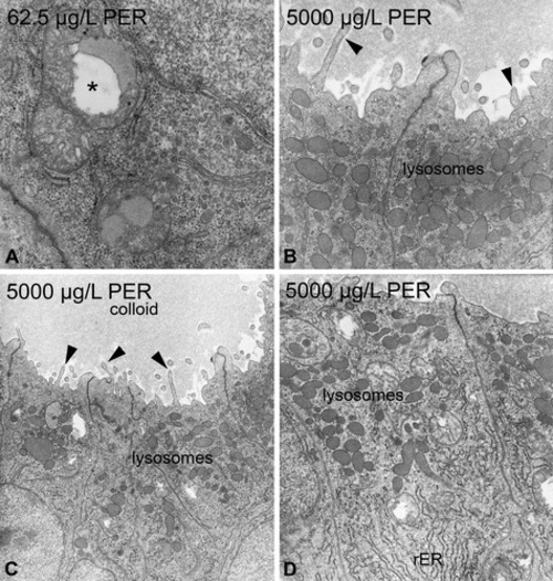
Perchlorate-induced ultrastructural alterations in zebrafish thyrocytes. At concentrations ≥62.5 µg/L, mitochondria are swollen and display irregular swellings of the intercristae space (*; A). At concentrations of 5,000 µg/L, a marked increase in lysosomes mostly located in the apical part of thyrocytes was visible (B, C, D). The rough endoplasmic reticulum showed moderate proliferation and some fenestration (D). The electron density of the colloid markedly decreased in higher concentration groups (C). Proliferations of microvilli are observable in concentrations ≥62.5 µg/L (▸; B, C). Magnification: A: 31,500×; B: 20,000×; C: 10,000×; D: 16,000×. |